
Page 138 - Haematologica Vol. 107 - September 2022
P. 138
ARTICLE - A mouse model of humanized type 2B VWD
S. Kanaji et al.
ligand engagement, elevated shear, and coagulation.19 The expression of GPVI on platelet surface was also decreased in VWF2Bhet hGPIba and VWF2Bhomo hGPIba mice compared to VWFhA1 hGPIba mice (Online Supplementary Figure S1B). Western blotting of VWF2Bhomo hGPIba mouse platelet lysate confirmed loss of full-length hGPIba, leaving C-terminal fragment after shedding (Online Supplementary Figure S1C). Interestingly, the amount of intracellular filamin A (FlnA) was decreased in VWF2Bhomo hGPIba mouse platelets com- pared to VWFhA1 hGPIba mouse platelets. The level of FlnA was also quantified by staining platelets intracellularly after fixation and permeabilization, followed by platelet sizecorrection(OnlineSupplementaryFigureS1D).Reduced FlnA content in VWF2Bhomo hGPIba mouse platelets was con- firmed by flow cytometry, indicating activation of m-calpain and enhanced FlnA degradation in VWF2Bhomo hGPIba mouse platelets.20,21 Functional defect of platelets in response to agonist stimulation has been reported in patients and mouse model of type 2B VWD.22-24 Thus, mouse platelets have been stimulated with PAR4 activating peptide (PAR4- AP) and fibrinogen binding was evaluated by flow cyto- metry. As expected, fibrinogen binding was impaired in VWF2Bhet hGPIba and VWF2Bhomo hGPIba mouse platelets compared to VWFhA1 hGPIba mouse platelets (Online Sup- plementary Figure S2). Defect in fibrinogen binding follow- ing agonist stimulation has also been previously reported in a mouse model of platelet-type VWD.25
Contribution of megakaryocyte/platelet versus endothelial cells to the pathophysiology of type 2B von Willebrand disease
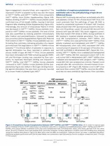
Plasma VWF is primarily derived from endothelial cells (EC), and platelets contain 10-15% of total blood VWF.1 Thus, one could postulate that the pathology of type 2B VWD is caused by endothelial synthesis of mutant VWF. Previous study showed that VWF spontaneously bound to the sur- face and intracellular canals of cultured MK derived from patients with type 2B VWD.6 This result suggests a possi- bility that mutant VWF binds to GPIba during synthesis in MK, leading to impaired platelet generation. Thus, recip- rocal BM transplantation between VWFhA1 hGPIba and VWF2Bhomo hGPIba mice was performed. VWFhA1 hGPIba mice lethally irradiated and transplated with VWF2Bhomo hGPIba BM hematopoietic stem cells (HSC) expressed VWF with normal human A1 sequence in EC and gain-of-function mu- tant VWF (p.V1316M) in MK/platelets (2BMK, Figure 3A). Con- versely, VWF2Bhomo hGPIba mice irradiated and transplanted with VWFhA1 hGPIba BM HSC expressed mutant VWF in EC and normal VWF in MK/platelets (2BEC). VWFhA1 hGPIba mice irradiated and transplanted with allogenic VWFhA1 hGPIba mouse BM HSC were prepared as controls. Platelet count analyzed after BM reconstitution showed marked throm- bocytopenia in 2BEC (Figure 3B). Platelet count in 2BMK mice was lower than control VWFhA1 hGPIba mice but the differ- ence did not reach statistical significance. Flow cytometry
AB
C
Figure 3. Crossed bone marrow transplantation between VWFhA1 hGPIbα and VWF2Bhomo hGPIbα mice. (A) In order to express type 2B von Willebarnd factor (VWF) selectively in endothelial cells (EC) or megakaryocytes[MK]/platelets, VWFhA1 hGPIba and VWF2Bhomo hGPIba mice were lethally irradiated (1,100 cGy) and transplanted with hematopoietic stem cells (HSC) derived from the other strain. (B and C) Platelet count and platelet flow cytometry analyses. Surface expression of GPIba and VWF binding were analyzed using the method described in Figure 2A after bone marrow (BM) reconstitution. Data are shown as mean ± standard deviation. Statistical analysis performed with the Kruskal-Wallis non-parametric test followed by Dunn’s multiple comparison test. *P<0.05, ****P<0.0001.
Haematologica | 107 September 2022
2137
