
Page 151 - 2022_02-Haematologica-web
P. 151
RHOA mutation analysis in early lymphoma diagnosis
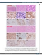
Figure 3. Histological and immunophenotypic findings in case 30. The first lymph node excision biopsy shows partial effacement of the lymph node architecture by polymorphous infiltrates, particularly B cells with plasmacytoid differentiation. A large proportion of B cells were EBER-positive and showed IG κ light chain restriction (not shown). The lymphoid follicles appear to be reactive and shows no apparent expansion of T follicular helper (TFH) cells, with only a few CD10-positive cells spilling out of the germinal center, consistent with pattern-1 histology of angioimmunoblastic T-cell lymphoma (AITL). The second lymph node biopsy shows effacement of the lymph node architecture by medium-sized atypical lymphoid cells with regressed follicles. There is a prominent proliferation of follicular dendritic cell meshworks and high endothelial venules with atypical lymphoid cells clustered in their vicinity. The atypical lymphoid cells are T cells expressing TFH markers, spilling out of the germinal center to the interfollicular region. EBER in situ hybridization shows only scattered positive cells.
haematologica | 2022; 107(2)
493
