
Page 149 - 2022_02-Haematologica-web
P. 149
RHOA mutation analysis in early lymphoma diagnosis
B
A
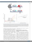
Figure 1. Detection of RHOA p.Gly17Val (c.50G>T) by real time polymerase chain reaction with peptide nucleic acid/locked nucleic acid probes. (A) Schematic illustration of the real time polymerase chain reaction (PCR) design. The total probe is used as an internal control to monitor the PCR performance. The peptide nucleic acid (PNA) clamp binds to the wild-type sequence, resists the 5’ nuclease activity of Taq DNA polymerase and thus blocks the wild-type allele from PCR ampli-
fication. This results in preferential amplification of the mutant allele which is detected specifically by the locked nucleic acid (LNA) mutant probe. (B) The PCR assay is highly sensitive, capable of detecting the RHOA mutation at a variant allele frequency (VAF) of 0.03% based on serial dilutions of an angioimmunoblastic T-cell lymphoma sample with known mutation allele frequency by next-generation sequencing (left panel). For simplicity, only the LNA mutant but not total probe signals are shown. The PCR assay shows a linear correlation among the serial dilutions (right panel).
the qPCR assay using serial dilutions of three purified DNA samples with known VAF of Gly17Val (c.50G>T) into tonsil DNA. The PNA-LNA qPCR was highly sensi- tive, capable of detecting the mutation at a VAF of 0.032% (Figure 1B). As expected, the qPCR was highly specific to Gly17Val (c.50G>T) and showed no detectable signal of the LNA mutant probe with the DNA samples harboring other RHOA mutations including Gly17Leu (c.49-50GG>TT) and Gly17Glu (c.50G>A), or tonsillar DNA (Online Supplementary Figure S1A).
As crude DNA preparations were routinely used for clonality analysis in our clinical diagnostic laboratories, we tested whether such crude DNA samples were amenable to the above PNA-LNA qPCR (Online Supplementary Figure S1C). Of the ten crude DNA sam- ples initially tested, nine yielded excellent amplification comparable with the results from purified DNA samples. We then used crude DNA preparations for the qPCR in the remaining investigations, with high quality results from 139 of the 144 samples investigated. The reason that the other five samples failed to support qPCR was
essentially the poor quality of the DNA, as evidenced by quality control PCR.
RHOA Gly17Val (c.50G>T) is detected in initial biopsies not diagnostic for lymphoma by conventional approaches
To confirm that the RHOA mutation was lymphoma- specific, not associated with reactive conditions prolifer- ations or other lymphoproliferative disorders with enriched T cells, we investigated 45 tissue biopsies including 27 from lymph nodes, for which T-cell clonali- ty analysis was requested during routine histological diagnosis, but showed no evidence of a T-cell lymphoma. These included 13 specimens showing paracortical T-cell expansion or enriched CD4+ T cells, and ten classic Hodgkin lymphomas with a prominent background of T cells. The qPCR was successful for all these specimens, as indicated by the total probe control, but they were all negative for the RHOA mutation. Similarly, we also showed absence of the RHOA mutation in peripheral blood lymphocytes (Online Supplementary Figure S1D)
haematologica | 2022; 107(2)
491
