
Page 150 - 2022_02-Haematologica-web
P. 150
R. Dobson et al.
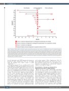
Figure 2. Analysis of RHOA (c.50G>T; p.Gly17Val) mutation by quantitative polymerase chain reaction in the RHOA-positive cases with longitudinal biopsies in patients with angioimmunoblastic T-cell lymphoma (only including cases with at least one biopsy classed as not diagnostic [“group B” or “group C”]). The diagnoses of these specimens were reviewed, and categorized into three groups: group A (filled circles): lymphoma diagnosed by histology and immunophenotype, further sup- ported by clonal TCR gene rearrangement; group B (half-filled circles): lymphoma not diagnostic by histology and immunophenotype alone, but ascertained by clonal TCR gene rearrangement; group C (open circles): lymphoma not diagnosed by combined analyses. Red (regardless of the symbol: including outline only, half-filled, and completely filled symbols) indicates RHOA p.Gly17Val mutant positive biopsy. Gray indicates RHOA p.Gly17Val status unknown. E denotes extranodal biopsies. *denotes lymph node excision biopsies and all others are core biopsy specimens. An open square denotes a diagnosis of classic Hodgkin lymphoma with no apparent evidence of angioimmunoblastic T-cell lymphoma (AITL). Case 38 lacks a final diagnosis as the patient died, indicated by a dashed line. Cases with only group A mul- tiple biopsies are not included in this figure.
from 16 individuals with CHIP (mutational information provided in Online Supplementary Table S2), in keeping with the findings from whole exome or panel sequencing.17,18
We then investigated 37 cases of AITL (n=35) or nodal PTCL-TFH (n=2) with unknown RHOA mutation status, of which 29 had multiple longitudinal biopsies (2-5) avail- able for RHOA analysis. RHOA mutation was seen in 23 cases, but was negative in the remaining 14 cases. Among the 23 cases with RHOA mutation, 19 cases had multiple biopsies. We reviewed the original histological diagnosis in each of these specimens and categorized them into three groups. Group A specimens (n=26) showed clear evidence of lymphoma by histology and immunopheno- type, further supported by clonal TCR rearrangement. Group B (n=6) had suspicious histological and immunophenotypic findings, although not entirely diag- nostic for lymphoma, which was ascertained by detec- tion of strong clonal TCR gene rearrangements. Finally, group C mutations (n=18) were not diagnostic for lym- phoma by combined histological, immunophenotypic
and clonality analyses (Online Supplementary Table S3). Interestingly, RHOA mutation was detected in each of these specimens, in the positive cases, including those not diagnostic for lymphoma by conventional integrated diagnostic investigations (Figure 2).
Characteristics of biopsies not diagnostic for lymphoma but positive for RHOA mutation
Of the 23 cases of AITL or PTCL-TFH with RHOA mutation, the initial biopsy was not diagnostic in ten cases including two with an excisional lymph node spec- imen. The time from the initial non-diagnostic biopsy to that establishing AITL or PTCL-TFH ranged from 0 to 26.5 months, with an average of 7.87 months. All these initial non-diagnostic biopsies showed polyclonal (n=4), weak clonal (n=5) or oligoclonal (n=2) TCR gene rearrangements or failed (n=1) by BIOMED-2 TRB and TRG clonality analysis. Seven of the initial non-diagnostic biopsies were also subjected to BIOMED-2 B-cell clonali- ty analysis and six showed a clonal IG gene rearrange- ment. Epstein-Barr virus (EBV)-encoded small RNA
492
haematologica | 2022; 107(2)
