
Page 153 - 2022_02-Haematologica-web
P. 153
RHOA mutation analysis in early lymphoma diagnosis
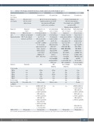
Table 1. Summary of histological, immunohistochemical, clonality analysis and genetic findings in case 7.
Time after the 1st biopsy Clinical presentation
Biopsy site
Histology
and Immuno-phenotype
First biopsy
Skin rash, fevers; All self-resolved with no therapy
Skin punch biopsy
Mild perivascular infiltrate, predominantly CD3+ T cells, admixed histiocytes and B cells
Second biopsy Third biopsy
11 months later 11.5 months later
Episode of non-specific symptoms; lymphadenopathy revealed by CT, but relatively low PET signal; opted for close surveillance.
Fourth biopsy Fifth biopsy
33 months later 35 months later
Slowing enlarging lymph node with increasing symptoms. Treated with 6 cycles of R-GCVP* and achieved CR; Ongoing remission in May 2020
Diagnosis
T-cell clonality TRG-A
TRG-B TRB-A TRB-B TRB-C
B-cell clonality BaseScope-ISH
for TRB-V5- Targeted sequencing
Panniculitis
n/a
n/a
n/a
n/a
n/a
n/a
Few positive cells
n/a
Lymph node core biopsy Predominant infiltrate
of small to medium sized T cells
with vasculocentric pattern, CD4+, some CD10+ and BCL6+ T cells possibly spilled outside B-cell follicle, expanded FDC meshworks; scattered large EBER+ B cells: CD30+, CD15 weak+
AITL
217 bp Poly
245 bp Poly
302 bp
n/a Diffuse positive
DNMT3A (VAF: 34%) (c.920C>T; p.P307L) TET2 (VAF: 8%) (c.3646C>T; p.R1216X) TET2 (VAF: 36%) (c.3781C>A; p.R1261S) TET2 (VAF: 3%) (c.3866G>T; p.C1289F) TET2 (VAF: 18%) (c.4947T>A; p.Y1649X) RHOA (VAF: 20%) (c. 50G>T; p.G17V) Strong positive
Left axillary lymph node core biopsy Very similar
to the 2nd biopsy. Predominantly
small to medium sized T cells associated with HEV proliferation, CD4+, PD1+, BCL6 variable+; Expanded FDC and pleomorphic infiltrate, scattered large B cells with HRS morphology CD30+, CD15 weak+ AITL lymphoma
217 bp Poly
245 bp Poly
Poly
n/a Diffuse positive
n/a
Right axillary lymph node core biopsy Lymph node
structure effaced by polymorphous infiltrate with scattered large atypical cells expressing CD30, CD20 (weak), CD79, PAX5, BCL6, MUM1, OCT2, BOB1, and EBER+,
but not CD15.
CD3 T cells:
largely CD4+, some ICOS+, occasional PD1+, but CD10– and CXCL13– Classic Hodgkin lymphoma
Poly Poly Poly Poly Poly Poly n/a
n/a
Right axillary lymph node core biopsy Normal structure largely effaced
by polymorphous infiltrate with prominent large atypical cells expressing CD30, CD20, CD79a, PAX5, MUM1,
BCL6(weak), and EBER+, but not CD15, CD10, CYCLIN D1, ALK and CD25.
No detectable T-cell abnormalities Classic Hodgkin
Poly Poly Poly Poly 187bp Poly Negative
DNMT3A (VAF:20%) (c.920C>T; p.P307L) TET2 (VAF: 4%) (c.3646C>T; p.R1216X) TET2 (VAF: 20%) (c.3781C>A, pR1261S) TET2 (VAF: 8%) (c.3866G>T; p.C1289F)
RHOA (VAF: 1%) (c.50G>T; p.G17V) Weak positive
RHOA c.50G>T
n/a:not available;R-GCVD:rituximab,gemcitabine,cyclophosphamide,vincristine,prednisolone;CR:complete remission;VAF:variant allele frequency.
Weak positive
Strong positive
Weak positive
haematologica | 2022; 107(2)
495
