
Page 284 - 2022_01-Haematologica-web
P. 284
A. Astori et al.
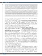
Figure 4. The RINF knockdown-induced phenotype is dependent on transforming growth factor b in human primary cells. (A) Transforming growth factor b (TGFβ) ligand and TGFBRI expression profiles were extracted from our proteomic analyses of cord blood-derived CD34+ cells (Gauthier et al.37). TGFB1 and TGFBRI were highly expressed at the Prog1-Prog2 stage (i.e. D1-D3 of erythropoietin [EPO] treatment), suggesting important autocrine signaling in early steps of erythroid maturation in our serum-free culture conditions. (B) Cord blood-derived CD34+ cells were transduced at day 5 of the amplification step, GFP-sorted (2 days after transduction), and EPO was added for 8 to 12 days, in the presence of TGFb inhibitor (SB431542, at 10 mM) or vehicle control (dimethylsulfoxide, DMSO). The histograms (left panel) represents fluorescence activated cell sorting (FACS) analyses of the whole cell suspension for CD49d and Band3 antigens (here at 10 days of EPO). In order to deter- mine whether the observed acceleration of maturation was statistically significant, Student t-tests were performed. Scattergrams (right panel) present one represen- tative experiment out of three separate experiments performed. (C) RINF (left panel) and phosphorylated SMAD2 (right panel) protein levels were estimated at 8 h and 72 h of treatment with EPO by western-blot analysis (see Methods), with the same kinetics as presented in Figure 2C (pool of 5 cord blood units). As expected, the specific band for phospho-SMAD2 (Ser465/467), indicated by an arrow, was not detected in the presence of TGFb inhibitor (SB431542, at 10 mM). P85 (PIK3R1) was used as a loading control (lower panel) from the same blot, and at a shorter exposure time than in Figure 2C.42 For each lane, protein extracted from 3x10⁵ cells was loaded. Band intensities were quantified with Multigauge software and the relative phospho-SMAD2 expression is shown (right panel). (D) TGFb (5 ng/mL) or vehicle control (DMSO) was added at 11 days of EPO and the proliferation monitored during 1 additional week, until day 18 of EPO. For both treated (in red) and untreated (in black) conditions, red blood cell expansion was lower for RINF-silenced cells. The kinetics shown corresponds to Donor#4 of Figure 2E (right panel). Cell culture growth is represented in population doublings (left panel) or cumulative cell number at day 18 of EPO (right panel). (E and F) The two donors (1 and 2) that seemed not sensitive to RINF knockdown in the absence of TGFb (Figure 2E) were treated with variable doses of TGFb1 (ranging from 0.1 to 2.5 ng/mL), and scattergrams (upper panel) represent FACS analyses of the whole cell suspension labeled for both GPA and CD71 antigens. The corresponding dose-effect curves are also shown (lower panel). One representative scattergram at the lowest concentration (0.1 ng/mL) of exogenous TGFb1 is shown. (F) A histogram representing GPA acquisition (upper panel) which is more advanced in RINF knockdown cells (here at day 6 of EPO) in the presence of low doses of TGFb1 (here at 0.1 ng/mL). Cell growth was also monitored, and the total number of red blood cells numerated at day 17 of EPO are presented in the histograms (lower panel).
and SMAD7, we next performed “rescue” experiments in K562 cells. We employed retroviral transduction to express SMAD7 ectopically (Figure 5D). Our data demon- strated that SMAD7 overexpression did not affect endogenous expression of RINF mRNA or protein (Figure 5E, F), but did reduce hemoglobinization (Figure 5G, lane 3), mimicking the effect of ectopic RINF (Online Supplementary Figure S5B). We did not detect any change in cell viability or proliferation in the absence of hemin. Importantly, under conditions in which SMAD7 was ectopically expressed, RINF knockdown did not result in acceleration of hemoglobinization (Figure 5G). Taken together, these data indicate that RINF binds the SMAD7 promoter directly to control its expression, and that SMAD7 is an effector downstream of RINF during ery- throid maturation.
RINF and SMAD7 mRNA expression are correlated in bone marrow from adult donors and patients with myelodysplastic syndrome
We investigated whether RINF and SMAD7 mRNA expression correlated in primary human CD34+ cells from healthy donors and donors with MDS. To this end, we performed qRT-PCR on CD34+ cells isolated from adult bone marrow donors (n=11) and found a highly signifi- cant correlation between RINF mRNA and SMAD7 mRNA (Pearson, rho=0.684, P=0.02) (Figure 6A). We con- firmed these data by analyzing an independent microar- ray dataset from Pellagatti et al.46 As shown in Figure 6B, RINF and SMAD7 mRNA levels correlated strongly in CD34+ cells isolated from healthy donors (Pearson rho=0.784, P<0.001, n=17). Interestingly, this correlation also reached statistical significance in CD34+ cells from MDS -5q patients (Pearson rho=0.603, P<0.001, n=47) but not in other MDS patients, i.e., without del(5q) or 5q-. We then compared the intensity of this correlation with the other 5q genes (n=48 genes) from the commonly deleted regions associated with high risk, and the CXXC5 probe sets were those correlating best with the SMAD7 probe set in both the healthy control group (rho=0.819, and rho=0.784) and the MDS patients’ cohort with del(5q) (Table 1) (Online Supplementary Figure S6A). Reinforcing the previously reported relevance of SMAD7 in MDS pathophysiology,33,34 SMAD7 expression was not only extinguished in CD34+ MDS samples compared to normal samples (in the microarray dataset from Gerstung et al.,47 (Online Supplementary Figure S6C), but patients
with the lowest SMAD7 expression had a significantly shorter overall survival (Online Supplementary Figure S6D).
RINF-silencing alters genome-wide hydroxymethylation
Since murine RINF has recently been described as a nec- essary platform for TET2 activity,15,48 we wondered whether RINF-silencing could affect the genome-wide 5hmC of human immature erythroid cells. As shown in Figure 6C, we observed a statistically significant loss of 5hmC detect- ed by immunofluorescence or flow cytometry (Figure 6D) in knockdown primary cells.
A RINF/SMAD7 axis controls erythroid maturation and red blood cell expansion in primary cells
We next investigated whether RINF knockdown altered SMAD7 expression in primary erythroid progenitors. As shown in Figure 7A, B, a 60-70% knockdown of RINF led to a statistically significant knockdown of SMAD7 (by 40-45%) in CD34+ cells isolated from cord blood (n=3 donors, paired Student t-test, P<0.001). Accordingly, a 30% knockdown of SMAD7 protein level was detected by immunofluorescence in primary erythroid progenitors (P<0.0001) (Figure 7C). For a direct demonstration of the relevance of this finely tuned regulation of SMAD7 medi- ated by RINF protein in primary HSPC, we next per- formed experiments in which we knocked down RINF and induced SMAD7 expression ectopically. To this end, we first transduced CD34+ cells with the doxycycline- inducible vector pInducer21/SMAD7-HA (Figure 7D), and then with the pTRIPUD3 vector that drives shRNA/RINF or shRNA/Control expression. Notably, in the presence of doxycycline, SMAD7 expression (Figure 7E, F) robustly prevented both the RINF knockdown-dependent acceler- ated maturation (Figure 7G) and reduction of RBC num- bers (Figure 7H).
Discussion
An increasing body of work suggests that RINF is involved in the maturation, function, or development of various cell types such as neural stem cells,21 endothelial cells,49 myoblasts,14 myofibroblasts,24 osteoblasts,23 as well as in kidney development,22 wound healing,24 and hair regrowth.50 Here, we report that the epigenetic factor RINF is a transcriptional regulator of SMAD7 which fine-tunes TGFb sensitivity of erythroid progenitors, findings that
276
haematologica | 2022; 107(1)
