
Page 100 - 2021_12-Haematologica-web
P. 100
J. Li et al.
marrow immune subsets of AML patients compared with healthy donors (Figure 2C). PVRIG and PVRL2 expression levels varied considerably amongst patients, but this did not correlate with AML subtype or the percentage of bone marrow blasts (Online Supplementary Figure S4).
In order to assess whether PVRIG blockade could enhance NK-cell killing of AML patient blasts, we co-cul- tured healthy donor PBMC with bone marrow from an AML patient with a high percentage (>90%) of PVRL2hiPVRlo AML blasts. In this context, NK cells showed significantly increased CD69 expression and degranulation with PVRIG or TIGIT blockade. In addition, combined PVRIG and TIGIT blockade was associated
with significantly higher NK-cell activation and degranu- lation (Figure 2G and H). Similar results were obtained for a further two AML patients tested (Figure 2I to L). This indicates that PVRIG blockade or combination PVRIG/TIGIT blockade could enhance NK-cell cytotoxic- ity against PVRL2+ tumor targets in AML patients.
PVRIG expression on natural killer cells is modulated by activation
In order to understand why PVRIG was not upregulated on AML patient NK cells, we explored the mechanisms reg- ulating NK cell PVRIG expression. In order to do this, we activated healthy donor NK cells for 24 hours via co-culture
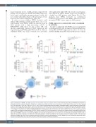
ABC
DEF
G
Figure 4. Modulation of DNAM-1 and TIGIT expression on natural killer cells upon activation. (A to C) T-cell immunoreceptor with Ig and ITIM domains (TIGIT) or (D to F) DNAX accessory molecule 1 (DNAM-1) expression on isolated natural killer cells after 24-hour culture with (A and D) K562 or KG1a cells (1:1 ratio); (Band E) 100 U/mL interleukin 2 (IL-2) and 10 ng/mL IL-12; or (C and F) with the indicated plate-bound antibodies. Percentage change in mean fluorescence intensity (MFI) relative to natural killer (NK) alone is shown, each point represents an individual donor (n=3), bars represent mean ± standard deviation (SD). Significance deter- mined by one-way ANOVA with Holm-Sidak’s multiple comparisons test (A, C, D and F) or Student’s t-test (B and E), not significant (ns) P>0.05, *P<0.05, **P<0.01, ***P<0.001, ****P<0.0001. (G) Model of PVRIG-TIGIT-DNAM-1 modulation upon NK-cell activation. Left: upon recognition of tumor cells expressing poliovirus receptor (PVR) and poliovirus receptor-related 2 (PVRL2), NK cells increase TIGIT but lose expression of both poliovirus receptor-related immunoglobulin domain-con- taining (PVRIG) and DNAM-1. This loss of DNAM-1 may result from a tumor-intrinsic mechanism of immune escape, whereby tumor cells expressing DNAM-1 ligands induce loss of DNAM-1 expression on immune cells. Right: upon activation via cytokines such as IL-2 and IL-12, or by stimulation of activating receptors such as CD16, NK cells decrease expression of PVRIG while increasing expression of TIGIT and DNAM-1. The increased expression of DNAM-1 relative to the decreased expression of PVRIG may serve to push the balance within the cell towards more activating signaling. In this way, PVRIG may act to constitutively dampen NK respon- siveness in the steady state and is lost upon activation to lower the activation threshold of the NK cell.
3120
haematologica | 2021; 106(12)
