
Page 98 - 2021_12-Haematologica-web
P. 98
J. Li et al.
blockade significantly decreased the NK:target ratio required to reach 10% KG1a lysis, while TIGIT blockade had only a minor effect (Figure 1B). PVRIG blockade also significantly increased NK-cell activation and degranula- tion, as measured by CD69 and CD107a staining respec- tively (Figure 1C and D). TIGIT blockade had minimal effect on both NK-cell activation and degranulation and combined PVRIG and TIGIT blockade showed no benefit over PVRIG blockade alone (Figure 1C and D). The acti- vating receptor DNAM-1 was important for recognition of targets, as blocking DNAM-1 significantly inhibited NK- cell activation and degranulation (Figure 1C ,D, H and I). KG1a target cell death was perforin-dependent, as it was completely blocked when free calcium was complexed with EGTA (Figure 1E).
PVRIG blockade was clearly more effective than TIGIT blockade for enhancing NK-cell responses against KG1a but did not enhance lysis of the breast cancer cell line
SKBR3. Rather, significantly more target cell death was observed with TIGIT blockade, or combined PVRIG and TIGIT blockade (Figure 1F). Pooled data from three donors suggested TIGIT blockade, but not PVRIG blockade, decreased the NK:target ratio required for 10% lysis, but the difference did not reach statistical significance (Figure 1G). TIGIT blockade significantly enhanced NK-cell acti- vation and degranulation, whereas PVRIG blockade had minimal effect on activation and a much smaller effect on degranulation (Figure 1H and I). Combined PVRIG and TIGIT blockade enhanced NK-cell activation and degran- ulation cytotoxicity even further, suggesting that PVRIG blockade can have an additive effect to TIGIT blockade (Figure 1H and I).
NK cells from all healthy donors tested expressed both PVRIG and TIGIT (Online Supplementary Figure S1). Although the levels of expression (particularly of TIGIT) varied, all donors were consistently more responsive to
ABC
DEF
GHI
JKL
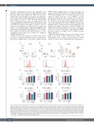
Figure 2. PVRIG and its ligand PVRL2 are expressed in acute myeloid leukemia patient bone marrow. Expression of (A) poliovirus receptor-related 2 (PVRL2) (B) poliovirus receptor (PVR) or (C) poliovirus receptor-related immunoglobulin domain-containing (PVRIG) on blasts or immune cell types in the bone marrow of acute myeloid leukemia (AML) patients (n=19-20) or healthy donors (n=13). Open triangles mark the patient shown in (D to F). Representative histograms of (D) PVRL2 and (E) PVR on AML blasts, or (F) PVRIG on natural killer (NK) cells in the bone marrow of an AML patient. Overlay histograms of test (red) and isotype control stains (grey) are shown. NK-cell expression of (G, I and K) CD69 and (H, J and L) CD107a after 4-hour co-culture of healthy donor peripheral blood mononuclear cells (PBMC) with AML patient bone marrow (8:1 E:T ratio) in the presence of the indicated blocking antibodies or isotype control antibody (mean ± standard deviation of triplicates, n= 3 patients). Significance was determined by one-way ANOVA with Holm-Sidak’s multiple comparisons test, not significant (ns) P>0.05, *P<0.05, **P<0.01, ***P<0.001, ****P<0.0001; MFI: mean fluorescence intensity.
3118
haematologica | 2021; 106(12)
