
Page 97 - 2021_12-Haematologica-web
P. 97
PVRIG blockade in acute myeloid leukemia
Results
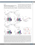
PVRIG blockade enhances natural killer cell killing of PVRL2hiPVRlo acute myeloid leukemia cells
In order to assess whether PVRIG blockade could enhance NK-cell responses against AML, we utilized the AML cell line KG1a. When co-cultured with healthy donor PBMC and PVRIG blocking antibody, a significant
increase in KG1a cell death was observed compared to the untreated control (Figure 1A). In contrast, TIGIT blocking antibody did not significantly enhance KG1a target cell lysis, and KG1a lysis in the presence of combined anti- PVRIG and anti-TIGIT was comparable to PVRIG block- ade alone (Figure 1A). In order to compare across donors with variable baseline killing, we calculated the NK:target ratio required for 10% KG1a lysis for each donor. PVRIG
AB
CDE
FG
HI
J
Figure 1. Blockade of PVRIG enhanced natural killer cell killing of tumor cell lines. (A) Percentage lysis of KG1a cells after 4-hour co-culture with peripheral blood mononuclear cells (PBMC) in the presence of anti-PVRIG, anti-TIGIT, anti-PVRIG + anti-TIGIT, or isotype antibodies, measured by 51Cr release assay. Representative data (mean ± standard deviation [SD] of triplicates) of four experiments shown. (B) Natural killer (NK):target ratio required to achieve 10% lysis, determined by non- linear regression of curves plotted as in (A). Each symbol represents an individual donor, n=4. NK-cell expression of (C) CD69 and (D) CD107a after 4-hour co-culture of PBMC with KG1a (8:1 E:T ratio) in the presence of the indicated blocking antibodies or isotype control antibody. Representative data (mean ± SD of triplicates) of two experiments is shown. (E) Percentage lysis of KG1a cells after 4-hour co-culture with PBMC in the presence of anti-PVRIG or isotype antibodies, with or without 4 mM EGTA. Representative data (mean ± SD of triplicates) of two experiments is shown. (F) Percentage lysis of SKBR3 cells after 4-hour co-culture with PBMC in the presence of anti-PVRIG, anti-TIGIT, anti-PVRIG + anti-TIGIT, or isotype antibodies, measured by 51Cr release assay. Representative data (mean ± SD of triplicates) of three experiments is shown. (G) NK:target ratio required to achieve 10% lysis, determined by non-linear regression of curves plotted as in (F). Each symbol repre- sents an individual donor (n=3). NK-cell expression of (H) CD69 and (I) CD107a after 4-hour co-culture of PBMC with SKBR3 (2:1 E:T ratio) in the presence of the indicated blocking antibodies or isotype control antibody. Representative data (mean ± SD of triplicates) of two experiments is shown. (J) Expression of PVRL2 and PVR (red histograms) on SKBR3 and KG1a cells compared with isotype control stain (grey histograms). NK:target ratios in (A, E and F) were calculated using % of NK cells in PBMC determined by flow cytometry. Significance was determined by repeated measures one-way ANOVA (A, B, F and G) or one-way ANOVA (C, D, H and I) with Holm-Sidak’s multiple comparisons test, not significant (ns) P>0.05, *P<0.05, **P<0.01, ***P<0.001, ****P<0.0001. PVRIG: poliovirus receptor-related immunoglobulin domain-containing; TIGIT: T- cell immunoreceptor with Ig and ITIM domains; MFI: mean fluorescence intensity.
haematologica | 2021; 106(12)
3117
