
Page 52 - 2021_09-Haematologica-web
P. 52
F. Barbaglio et al.
unmodified control cells (MEC1-CNTR) or MEC1-HS1KD cells with HS5 stromal cells as described above (Figure 1A) and observed that, outside the scaffolds, there were signifi- cantly fewer MEC1-HS1KD cells than MEC1-CNTR cells (P=0.0013) (Figure 1D). We then used quantitative real-time polymerase chain reaction (RT-qPCR) analysis to quantify the expression of HS1 in MEC1-CNTR cells cultured in 3D and found that HS1 was downregulated in MEC1 cells retained inside the scaffold compared to its level in the cells outside the scaffold (n=3 replicates; P=0.028) (Figure 1E).
Collectively, these findings indicate that our 3D system can reliably reproduce the BM-CLL interactions occurring in vivo and further underscore the relevance of HS1 downreg- ulation as a putative mechanism associated with CLL cell retention in the BM microenvironment, as previously sug- gested in mouse models.9
The bone marrow microenvironment regulates HS1 expression in primary chronic lymphocytic leukemia cells in 3D co-culture
We then co-cultured primary leukemic CLL cells isolated from the PB of six patients with CLL in the Spongostan scaffolds in the presence and absence of HS5 cells, as repre- sented in Figure 1A. We also confirmed for primary cells that stromal HS5 cells are needed to efficiently retain pri- mary CLL cells in the scaffolds (Figure 2A).
To further validate our model for CLL, we confirmed, by flow cytometry, the ability of CLL cells to retain the surface expression of their lineage markers CD19/CD5 throughout the whole culture period both inside and outside the scaf- folds (Online Supplementary Figure S1A).
We then quantified the expression of CXCR4 by RT- qPCR and flow cytometry in CLL cells recovered from inside and outside the scaffolds after 3 days of co-culture with HS5 cells and found that CXCR4 was downregulated in the cells retained inside the scaffold (n=8, P=0.015 Figure 2B; n= 3, P=0.03, Online Supplementary Figure 1C), mimick- ing the in vivo finding of CXCR4 downregulation in response to the binding of its cognate ligand CXCL12 (SDF- 1α).21 Next, we focused again on the HS1 gene and, similar to the findings in MEC1 cells (Figure 1E), we observed a sig-
nificant downregulation of HS1 in the CLL fraction inside the scaffolds as compared to the outside fraction (n=8 repli- cates, P=0.005) (Figure 2C). These findings raised the ques- tion of whether the expression of HS1 reflected a direct influence of the BM microenvironment or, conversely, a fraction of CLL cells constitutively expressing low levels of HS1 might preferentially home to the BM. In order to answer this question, we co-cultured primary CLL cells iso- lated from the PB of ten patients with the stromal cell line HS5 in two-deimensional (2D) monolayers, and evaluated the expression of HS1 by RT-qPCR. HS1 expression was significantly downregulated after 24 h of co-culture (n=15, P<0.0001) (Figure 2D). We then co-cultured CLL primary cells from the PB of eight patients with HS5 cells in the pres- ence and absence of a trans-well (1 μm pore) to avoid direct tumor-stroma contact. HS1 expression was not downregu- lated in the presence of the trans-well (Figure 2E), suggest- ing that its regulation requires direct contact between leukemic cells and the BM stromal microenvironment.
HS1 is heterogeneously expressed in chronic lymphocytic leukemia tissues
The results described above suggest that HS1 might be differentially expressed in CLL depending on the tissue in which the leukemic cells are located. To assess whether our 3D model recapitulates what occurs in vivo, we compared HS1 expression in primary human CLL cells from PB and BM. Using RT-qPCR, we observed that HS1 expression was significantly downregulated in CLL cells isolated from the BM as compared with its expression in paired samples iso- lated from the PB (n=9, P=0.0498) (Figure 3A). In the same cohort of patients, we also confirmed that CXCR4 expres- sion was downregulated in the BM as compared to the PB (n=10, P=0.003) (Online Supplementary Figure S1B). When we quantified HS1 expression at a single-cell level in CLL cells isolated from paired PB and BM samples using the Image Stream instrument,22 we observed that the majority of CLL cells in the PB strongly expressed HS1, while in the BM a fraction of CLL cells were HS1 negative (n=4, P=0.0187) (Figure 3B). Accordingly, immunohistochemistry performed on BM (n=4 patients analyzed) revealed a het-
A
B
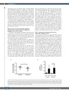
Figure 5. HS1 activation following ibrutinib treatment. (A) Dot plot showing the number of chronic lymphocytic leukemia (CLL) cells mobilized outside the scaffold after ibrutinib treatment and fractionated according to HS1 activation. CLL cells with active HS1 were mobilized more efficiently from the scaffolds compared to those with inactive HS1 (*P=0.04). (B) The graph shows the densitometric analysis of the active HS1-Y378 phosphorylated form in CLL cells retained inside the scaffold or recovered from outside the scaffolds with or without ibrutinib treatment. The CLL cells mobilized and recovered outside the scaffold show significantly higher HS1 activation than that of the cells retained inside the scaffold (**P=0.0035).
2340
haematologica | 2021; 106(9)
