
Page 51 - 2021_09-Haematologica-web
P. 51
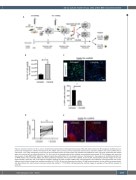
3D co-culture model of CLL cells within BM microenvironment
A
BC
DE
Figure 4. Ibrutinib treatment in 3D co-culture. (A) Schematic representation of the experimental setup: HS5 cells were seeded into 3D Spongostan scaffolds and cul- tured under microgravity in a RCCSTM bioreactor for 24 h. MEC1 cells or primary chronic lymphocytic leukemia (CLL) cells were added to the same scaffold and co- cultured for 72 h under microgravity. At the end of the incubation period, the supernatant was collected and depleted of cells. The same supernatant was added again to the culture with or without ibrutinib for 5 h. At the end of the incubation period the scaffolds were collected and analyzed. (B) The histogram plot shows the total number of cells (MEC1-GFP+ HS5) that migrated outside the scaffold after 72 h of dynamic culture in the bioreactor in the presence of 10 μM ibrutinib (for 5 h) or RPMI medium only (untreated). MEC1 cells were significantly mobilized (*P=0.02) from the scaffolds. (C) On the left, representative confocal images of the scaf- folds analyzed in panel (B), and on the right the histogram showing the mean number of MEC1 GFP+ cells quantified in the scaffold by counting the GFP+ cells in four different stacks for both treated and untreated conditions (P=0.002). (D) Line plot illustrating the effect of 10 μM ibrutinib treatment on the mobility of primary periph- eral blood-derived CLL cells that were recovered outside the scaffold (***P=0.0005). (E) Representative confocal images of examples of the scaffolds analyzed in panel (D).
haematologica | 2021; 106(9)
2339
