
Page 53 - 2021_09-Haematologica-web
P. 53
3D co-culture model of CLL cells within BM microenvironment
erogeneous pattern of HS1 expression among the patients (Figure 3C). This finding confirms that our 3D model can reliably reproduce the native BM microenvironment and provide significant insights into CLL cells in the tissues.
Chronic lymphocytic leukemia cells are mobilized from the scaffolds following exposure to ibrutinib
On the basis of the evidence presented here, HS1 appears to be involved in CLL cell compartmentalization, prompt- ing the question of whether it could also be involved in the process of CLL cell mobilization from the tissues. In order to address this point, we exploited our 3D model, as described in Figure 4A, and evaluated whether the cytoskeletal activity of HS1 also plays a role in the CLL cell mobilization promoted by the BTK inhibitor ibrutinib.16 MEC1 cells co-cultured with HS5 cells within scaffolds
were efficiently mobilized upon 5 h of treatment with ibru- tinib, as shown by both the number of MEC1 cells recov- ered in the medium (Figure 4B) and by the confocal images of the scaffolds, which exhibited significantly fewer GFP- tagged MEC1 cells in the untreated condition (P=0.002) (Figure 4C, Online Supplementary Movies 1-2). Of note, HS5 cells were not mobilized by the drug (Online Supplementary Figure S2A).
We next studied the response of primary CLL samples (n=21) to ibrutinib and found that the number of cells out- side the scaffolds was significantly higher upon drug treat- ment than the number in untreated samples (n=21, P=0.0005) (Figure 4D). Consistent with this, confocal microscopy analysis performed on the scaffolds showed that they were depopulated of CLL cells after incubation with ibrutinib (Figure 4E).
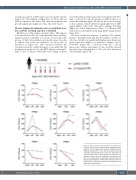
Figure 6. HS1 activation and expression in patients dur- ing ibrutinib treatment. Top left panel: schematic repre- sentation of the experimental protocol. Peripheral blood (PB) was collected at 1, 2, 3, 4, 8 and 12 weeks after drug treatment, PB lymphocyte (LY) count was determined at each time-point with a hemocytometer and chronic lym- phocytic leukemia cells were isolated and stored frozen at -80°C. For each patient, we plotted the western blot den- sitometry quantification of HS1-Y378 and the number of lymphocytes in the PB. The values are normalized to the basal level (n=8 patients).
haematologica | 2021; 106(9)
2341
