
Page 54 - 2021_09-Haematologica-web
P. 54
F. Barbaglio et al.
As expected, the expression levels of HS1 of cells moving out of the scaffolds decreased while they remained lower in CLL cells inside the scaffold in both untreated and treated (ibritunib) settings (Online Supplementary Figure S2B). Conversely, CXCR4 expression on CLL cells inside the scaf- fold decreased following exposure to ibrutinib, confirming previous in vivo results from Chen et al.17 (P=0.03) (Online Supplementary Figure S2C). In parallel, we observed that CXCL12 levels in the medium did not change significantly during the drug treatment (Online Supplementary Figure S2D).
Cells with inactive HS1 are less efficiently mobilized following ibrutinib treatment
To elucidate the mechanism underlying CLL mobiliza- tion, we evaluated whether HS1-mediated cytoskeletal rearrangements might be involved. We have previously shown that HS1 activation differs among patients with CLL and is associated with the clinical course (active HS1 is asso- ciated with a favorable prognosis while inactive HS1 is asso- ciated with an adverse prognosis).10 CLL cells with active HS1 (carrying HS1 phosphorylated in Y378, as determined by western blot analysis; data not shown) show efficient cytoskeletalfunctionality,whileCLLcellswithinactiveHS1 (not phosphorylated in HS1-Y378) display reduced cytoskeletal activity associated with a higher propensity to accumulate within the BM microenvironment.10 We detect- ed that both CLL cells with active HS1 and those with inac- tive HS1 were capable of homing to the scaffolds without significant differences (data not shown). Interestingly, we observed that the CLL cells released from the scaffolds upon exposure to ibrutinib were enriched for those with active HS1 (10 cases) as compared to those with inactive HS1 (10 cases) (P=0.04) (Figure 5A). Similarly, when we studied, in the same co-culture model, HS1 activation, i.e. the levels of HS1-Y378 determined by western blot, in primary CLL cells (n=7) exposed or not to ibrutinib, we observed that the lev- els of HS1-Y378 were higher in the cells released into the supernatant than in those retained inside the scaffolds (n=7, P=0.0035) (Figure 5B). These results demonstrate that more aggressive CLL cells (i.e., those with inactive HS1) are less efficiently mobilized from the BM surrogate scaffold and confirm that the segregation of CLL cells between the two compartments is not random but rather affected by the drug that, in turn, mediates changes in HS1 activation. Arguably, therefore, our 3D model may discriminate between cases that will respond more robustly to the CLL cell-mobilizing effect of ibrutinib.
Chronic lymphocytic leukemia cells mobilized into the peripheral blood by ibrutinib show active HS1 during the first weeks of treatment
Finally, we analyzed HS1 activation in CLL cells of patients (n=8) under ibrutinib treatment for different peri- ods (from week 1 to week 12) and correlated it with the lymphocyte count in the PB at the different time-points. In accordance with the results obtained in our 3D model (Figure 5B), we observed that PB CLL cells from six of the eight patients displayed HS1 activation during the first weeks (weeks 1, 2, and 3) of treatment as compared to the basal level (Figure 6A), in parallel with an increase in lym- phocyte count in the PB. In contrast, peripheral lymphocy- tosis was not seen in the two patients in whom HS1 did not undergo activation. In parallel, we analyzed HS1 expression in CLL cells by both western blotting and RT-qPCR and
found that HS1 expression increased during the first weeks of treatment in all patients analyzed either at the protein or at the gene level; however, we could not find a correlation with circulating lymphocyte count (Online Supplementary Figure S3A). Collectively, these results indicate that our 3D model can mirror the events occurring in vivo during ibruti- nib therapy.
Discussion
The clinical scenario of CLL is rapidly changing, in partic- ular thanks to new targeted therapies,23 although the dis- ease is still incurable. CLL is strongly influenced by the tis- sue microenvironment, as evidenced by the fact that circu- lating CLL cells are more sensitive to drug-induced apopto- sis, suggesting that a supportive microenvironment is nec- essary for the survival of leukemic cells. This has encour- aged the development of mobilizing agents24 and points to the key role of the cytoskeleton in recirculation and accu- mulation of CLL cells in different tissues.
A key prerequisite for investigating the mechanisms underlying human CLL cell homing and mobilization is the capability to reliably and accurately reproduce a native CLL tissue microenvironment in vitro, which is so far unavailable. Our previous studies pointed to the importance of the BM microenvironment, showing spe- cific homing of aggressive CLL (with inactive HS1) to this site in vivo in mouse models.9 For this reason, following our previously published experience from the analysis of multiple myeloma cell survival and response to borte-
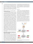
Figure 7. Schematic summary of the presented model. CLL: chronic lymphocyt- ic leukemia; BM: bone marrow.
2342
haematologica | 2021; 106(9)
