
Page 49 - 2021_09-Haematologica-web
P. 49
3D co-culture model of CLL cells within BM microenvironment
dures, the operational conditions of the RCCSTM were set and con- stantly monitored in order to keep the samples in a “free fall” con- dition, which minimizes sedimentation of the scaffold while max- imizing mass transfer and cell viability for the extended culture period.
Ibrutinib treatment in the bioreactor
After 72 h of 3D dynamic culture in the bioreactor, supernatants were withdrawn from the vessels and centrifuged at 1,500 rpm for 5 min. Recovered cells were counted. Clarified supernatants were put into the vessels again, with or without 10 μM ibrutinib. We compared two different concentrations of ibrutinib (1 and 10 μM) to exclude a possible role of cell apotpotosis in the mobilization from the scaffold, due to the possible increased toxicity of ibruti- nib at the higher concentration, and did not observe any signifi- cant differences (Online Supplementary Figure S2E). Cultures were stopped after 5 h of treatment and cells in the supernatants and in the scaffolds were recovered and submitted to the above men- tioned analysis (see Online Supplementary Methods).
Results
Microenvironmental elements are required to establish a 3D culture bone marrow model for chronic lymphocytic leukemia
We customized a new 3D co-culture model, previously validated by our group for myeloma cell survival19 to recre- ate CLL and BM-stromal cell interactions inside a scaffold kept in culture in a rotating bioreactor (Figure 1A). We selected scaffolds made of Spongostan, which has an ultra- structure similar to the trabecular structure of BM, also because of their superior performance in supporting CLL cell retention compared to either gelatin or collagen-coated beads (data not shown). To set the optimal experimental con- ditions, we first defined the best ratio of cellular compo- nents and the most appropriate co-culture medium for sup- porting cell viability (see Methods). The scaffolds were sequentially populated with the human BM-derived stro- mal cell line HS5 and the CLL cell line MEC1. Scaffolds
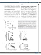
A
Figure 2. HS1 expression is regulated by the stromal bone marrow microenvironment. (A) The graph shows the total number of cells recovered in the medium outside the scaffold after 72 h of dynamic culture in the bioreactor. For the experiments with primary chronic lymphocytic leukemia (CLL) cells, control samples were run using CLL cells alone and HS5 cells alone under the same culture conditions. CLL cells were retained inside the scaffolds only in the presence of HS5 (**P=0.0047). (B, C) The line plots show CXCR4 and HS1 expression, respec- tively, as determined by quantitative real-time polymerase chain reaction (RT-qPCR) in primary CLL cells collected from inside the scaffolds or from the outside medium. CLL cells retained inside the scaffold expressed significantly lower levels of CXCR4 (n=8; *P=0.015) and HS1 (**P=0.005). (D) Line plot illustrating the down-regulation of HS1 expression in CLL cells iso- lated from peripheral blood when they were in direct co-culture with HS5 cells, as determined by RT-qPCR. (****P<0.0001). (E) Line plot illustrating the HS1 expression in CLL cells isolated from peripheral blood when they were cultured in a 1 μm pore trans-well without direct contact with HS5 cells.
BC
D
E
haematologica | 2021; 106(9)
2337
