
Page 52 - 2021_06-Haematologica-web
P. 52
G. Forlani et al.
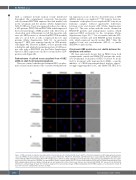
with a confluent dot-like fashion evenly distributed throughout this compartment; conversely Tax-1-positve cells of patients PH171206 expressed the viral marker both in the cytoplasm and the nucleus (Online Supplementary Table S1). Of note, Tax-1 was expressed in those two chron- ic ATL patients, PH170706 and PH171206, which expressed the lower percentage of HBZ positive cells. Moreover, as observed in acute ATL patients, not all Tax-1-positive cells were also HBZ-positive, as observed in PH170706 in which only 1% out of 6.4% of cells co-expressed the two viral proteins (Online Supplementary Table S1). As previously reported,26,27 HBZ was expressed mostly in CD4-positive cells (Figure 4A). However in PBMC of those patients with a distinctive and relatively normal proportion of CD8-posi- tive cells, such as PH170918 (see the Online Supplementary Figure S1), HBZ expression could also be found in few CD8- positive cells (Figure 4B).
Predominance of spliced versus unspliced form of HBZ mRNA in adult T-cell leukemia-lymphoma
Then we verified whether predominant HBZ cytoplas- mic localization in leukemic cells correlated with preferen-
A
tial expression of one of the two described forms of HBZ mRNA, spliced versus unspliced.37,38 We found a clear pre- dominance of the spliced versus unspliced HBZ form in our leukemic samples without appreciable differences between acute and chronic ATL (Online Supplementary Figure S3). This was in line with the results obtained in HAM/TSP patients and asymptomatic carriers, which expressed HBZ exclusively in the cytoplasm (Online Supplementary Figure S3),26,27 but at variance with the ATL- 2 leukemic cell line and with PH1505 patient leukemic cells, which expressed mostly nuclear HBZ.27 Here the unpliced form was predominant (ATL-2) or similar (PH1504) to spliced form.
Cytoplasmic HBZ protein does not shuttle between the cytoplasm and nucleus
We have previously shown that in PBMC from both asymptomatic carriers and HAM/TSP patients the exclu- sive cytoplasmic localization of HBZ could not be modi- fied by treatment with Leptomycin B (LMB), a specific inhibitor of CRM1/exportin-mediated nuclear export,39 strongly suggesting that in AC and HAM/TSP, HBZ does
B
Figure 3. HBZ subcellular localization in peripheral blood mononuclear cells of patients with chronic adult T-cell leukemia-lymphoma. (A) Peripheral blood mononu- clear cells (PBMC) of representative chromic leukemia patients PH150610, PH170706, PH170918 and PH171206 were stained with the 4D4-F3 anti-HBZ mono- clonal antibody (mAb) followed by Alexa Fluor 546-conjugated goat anti-mouse IgG1 antibody (red) and analyzed by confocal microscopy. DRAQ5 fluorescence probe was used to detect the nucleus. (B) PBMC of representative chronic leukemia patient PH170706 was stained with the A51-2 anti-Tax-1 mAb followed by Alexa Fluor 488-conjugated goat-anti-mouse IgG2a antibody (green) and analyzed by confocal microscopy. Nucleus was stained with DRAQ5. DIC represents the differential interference contrast image. At least 300 cells were analyzed. One representative image of HBZ staining derived from PBMC samples of each patient is shown. At least 300 cells were analyzed. All scale bars are 5mm.
2080
haematologica | 2021; 106(8)
