
Page 50 - 2021_06-Haematologica-web
P. 50
G. Forlani et al.
found in a minority of the HBZ-positive cells with the exception of patient PH140126 in which all HBZ-positive cells expressed the viral protein in both cellular compart- ments. Nevertheless, even in the case of PH140126, the nuclear localization was clearly under-represented with respect to the cytoplasmic localization (Figure 1A). In acute ATL, the proviral load (PVL) was generally higher than chronic ATL and did not strictly correlate with the percentage of HBZ-positive cells (Online Supplementary Table S1), even though the limited number of patients ana- lyzed does not allow to draw a conclusion in terms of sta- tistical significance. Interestingly, PVL values exceeded one copy of viral genome per cell in three of four patients, suggesting multiple integrations per single cell, as also demonstrated by recent molecular studies at a clonal level.36
In order to compare expression and subcellular localiza- tion of HBZ with those of Tax-1, a similar analysis was performed. Three of four acute ATL, namely PH140126, PH1612N07 and PH160822 showed expression of Tax-1 in 65%, 8,5% and 43% of cells, respectively, while
PH131213 ATL were negative for Tax-1 (Online Supplementary Table S1). It is interesting to note that the two patients expressing the highest number of Tax-1 pos- itive cells (PH140126 and PH160822), were the same expressing the highest number of HBZ-positive cells.
As found for HBZ, Tax-1 subcellular localization was predominantly seen in the cytoplasmic compartment (Figure 2A and B; Online Supplementary Table S1). In assess- able samples, most, but not all, Tax-1 positive cells were positive also for HBZ (Online Supplementary Table S1); in these cases HBZ and Tax-1 were mainly co-localized in the cytoplasmic compartment (Figure 2C).
HBZ cytoplasmic localization is found also in chronic adult T-cell leukemia-lymphoma
Four patients, clinically defined as chronic ATL, were analyzed. We first assessed the PBMC cell surface pheno- type of these patients; as expected it was quite distinct from the phenotype observed in acute ATL. CD3 marker identifying all T cells, was clearly assessable in the major- ity of the cells in all patients analyzed with a noticeable
B
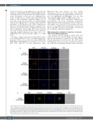
A
Figure 1. HBZ subcellular localization in peripheral blood mononuclear cells of patients with acute adult T-cell leukemia-lymphoma. (A) Peripheral blood mononu- clear cells (PBMC) from PH131213, PH1401263, PH160822 and PH1612N07 patients were stained with the 4D4-F3 anti-HBZ monoclonal antibody (mAb) followed by Alexa Fluor 546-conjugated goat anti-mouse IgG1 antibody (red) and analyzed by confocal microscopy; DRAQ5 was used to detect the nucleus. (B). Specific coun- terstaining of cytoplasmic compartment in PH1612N07 patient’s PBMC was performed by using anti-vimentin rabbit polyclonal antibody followed by goat anti-rabbit IgG conjugated to Alexa Fluor 488 (green). DIC represents the differential interference contrast image. At least 200 cells were analyzed. A representative image of HBZ staining is shown of each patient. All scale bars are 5mm.
2078
haematologica | 2021; 106(8)
