
Page 54 - 2021_06-Haematologica-web
P. 54
G. Forlani et al.
in the nucleus (Online Supplementary Figure S5B, PH160822 and PH170706).
Discussion
The nuclear localization of HBZ in ATL has been largely inferred from studies describing the many functions that this protein exerts directly on cellular gene promoters of infected cells or by interacting with a large number of host transcription factors in the nucleus.6,43-46 Recent investiga- tions conducted by confocal microscopy and biochemical approaches focused on endogenous HBZ protein were consistent with the idea that leukemic cells from HTLV-1- infected ATL patients express the viral protein in the nucleus.29 These findings, in association with elegant stud- ies in HBZ-transgenic animal models,15 gave strong sup- port to the idea that HBZ nuclear localization is instru- mental for, or at least strongly associated with, the main- tenance of the leukemic state. On the other hand, more recent and precise immune localization studies have clear- ly indicated that HBZ expression is always and only con- fined to the cytoplasm in HTLV-1 AC and in HAM/TSP patients in contrast to Tax-1 localization that can be found in both compartments.26,27 Since AC and more rarely HAM/TSP states may evolve to the leukemic state, it could be, thus, realistic to think that HBZ translocation from the cytoplasm to the nucleus is associated with the genesis of the neoplastic process leading to ATL. In order to verify this hypothesis it was instrumental to demon- strate that stages of leukemic transformation could indeed show evidence of cytoplasmic-only or cytoplasmic- nuclear localization of endogenous HBZ protein. In this study, we have demonstrated that indeed leukemic cells from ATL patients could be found that express HBZ in the cytoplasm with high frequency. Apparently, therefore, we filled the gap with previous studies and showed the unidi-
rectional subcellular translocation from the cytoplasm to the nucleus of HBZ during oncogenic transformation. Moreover, HBZ was expressed not only in CD4+ T cells, the preferential target of the HTLV-1 retrovirus, but also in a very small percentage of CD8+ T cells indicating that CD8+ T cells can be infected by HTLV-1, confirming our previous observations,26,27 and persist in ATL patients.
Several findings reported in this study require further investiagtions to be framed in the context of unidirection- al cytoplasm-to-nucleus localization of HBZ in ATL.
A rather unexpected and important finding was that none of the eight patients, either acute or chronic ATL, analyzed in this study had cells expressing HBZ exclusively in the nucleus. As a partial explanation, we can adduce that most of the previous studies describing endogenous HBZ localization were carried out in leukemic cell lines possibly representative of highly selected phenotypes in culture, while only three patients were described with an exclusive nuclear localization by a similar confocal microscopy approach.27,29 In this context, it is important to stress that previously analyzed ATL patients and those described in this study did not differ in terms of geographic origin or ethnic group being all afro caribbeans but one (PH150610).
A second important finding relates to the evidence, again common to both acute and chronic ATL, that in cells expressing HBZ in both the cytoplasm and nucleus, the cytoplasmic fraction was always consistently higher than the nuclear fraction. Actually, in two ATL chronic patient (PH150610 and PH170706), HBZ was only observed in the cytoplasm. A relative exception was represented by the acute ATL patient PH140126 displaying the highest per- centage of HBZ-positive cells (83%) all of which were positive for both cytoplasmic and nuclear localization. The unprecedented HBZ cytoplasmic localization in leukemic patients found in this study was paralleled by the known more abundant expression of the spliced versus
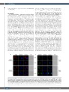
AB
Figure 5. Cytoplasmic HBZ is retained in this compartment and does not shuttle to the nucleus. (A) Peripheral blood mononuclear cells (PBMC) of representative acute PH140126 and chronic PH170918, were either treated (+LMB, bottom panels) or not treated (-LMB, top panels) with Leptomycin B (LMB), an inhibitor CRM1/exportin-mediated nuclear export, before fixing and stained with the anti-HBZ 4D4-F3 monoclonal antibody (mAb) followed by Alexa Fluor 546-conjugated goat anti-mouse IgG1 antibody (red) and analyzed by confocal microscopy. (B) As control of inhibition of nuclear export by LMB, treated (+LMB) and untreated (-LMB) cells were fixed and stained with antibodies against p65/RelA followed by Alexa Fluor 546-conjugated goat-anti-rabbit secondary antibody (red) and analyzed by con- focal microscopy. One representative image on the field and an enlargement of this are shown. Nucleus was stained with DRAQ5 (blue). DIC represents the differ- ential interference contrast image. One representative image is shown for each sample. At least 300 cells were analyzed. All scale bars are 5mm.
2082
haematologica | 2021; 106(8)
