
Page 138 - 2021_05-Haematologica-web
P. 138
M. Ackermann et al.
approved by the local ethical committee of Hannover Medical School (No.: 2127-2014 and 1303-2012).
Results
Interleukin 3 allows the generation of immature myeloid cells from induced pluripotent stem cell-derived hemanoids
We recently established a hematopoietic differentiation system that facilitates the generation of different mature myeloid cells from human iPSC via the generation of 3D complexes, which we refer to as “hemanoids” (Figure 1A).
These complexes are, depending on the cytokine adminis- tration, able to produce different myeloid cell types such as macrophages and granulocytes13 or erythrocytes24 for several months, suggesting the development and mainte- nance of a hematopoietic progenitor population. While we have intensively employed this approach for disease modeling studies and cell-based therapies,25-29 we here sought to utilize these organoid-like 3D complexes to study human embryonic hematopoietic development in vitro.
In order to elucidate the effect of IL-3 on the hematopoi- etic differentiation of hiPSC, we first evaluated the effect of cytokine withdrawal in our hemanoid-based differenti-
AB
C
E
D
F
G
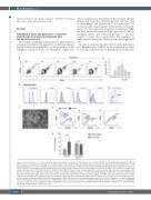
Figure 3. Characterization of hemato-endothelial progenitors within hemanoids. (A and B) Frequency of CD34+/CD144+ hemato-endothelial progenitors (HEP) at different days of hematopoietic specification. (A) Representative flow cytometry data (percentage of gated population is indicated) and (B) quantification of n=4, mean±standard error of the mean (SEM). (C) Detailed surface marker phenotype of HEP derived from hemanoids day 4 analyzed by flow cytometry (pre-gated on CD34+/CD144+ cells, histogram overlay: unstained grey filled, surface marker expression blue, representative data from n=3, approx. 1,000 HEP were analyzed per sample). (D) Expression of the IL3Ra/CD123 on CD34+/CD144+ HEP (histogram overlay: unstained grey filled, surface marker expression blue, representative data from n=3, approx. 1,000 HEP were analyzed per sample, percentage of gated population is indicated). (E) Morphology of HEP after 1 week in endothelial to hematopoietic transition (EHT) culture (scale bars: 100 mm and 200 mm, respectively). (F) Flow cytometry analysis of CD144, CD43 and CD45 expression on CD34+ cells after 1 week EHT culture (unstained: grey, surfacemarker expression: blue, representative data from n=2, approx. 10,000 CD34+ cells were analyzed per sam- ple, percentage of gated population is indicated, pre-gating on CD34 is shown in the Online Supplementary Figure S3C). (G) Left: Frequency of colony forming units (CFU) of iPSC-derived CD34+ cells after 1 week of EHT culture and cord blood-derived CD34+ cells (n=4, two biological and two technical replicates, mean±SEM). *P<0.05, **P<0.01, ns: not significant; statistical significance was assessed using (B) One-way ANOVA with Dunnetts multiple comparison test and (G) two-way ANOVA with Sidak's multiple comparisons test.)
1358
haematologica | 2021; 106(5)
