
Page 140 - 2021_05-Haematologica-web
P. 140
M. Ackermann et al.
expressed in (hemogenic) endothelium was noted in early phases of differentiation (EB and hemanoid day 4 to day 8), whereas expression of the hematopoietic TF TAL1 pro- gressively increased during the differentiation process (Figure 2C). In line with these results, we observed a strong downregulation of the pluripotency-associated sur- face antigen TRA-1-60 during the first days of differentia- tion. Moreover, expression of CD144 and CD34, both sur- face antigens expressed on (hemogenic) endothelium, peaked in hemanoids at day 8 of differentiation. The emergence of the first hematopoietic cells characterized by expression of CD43 and CD45 was observed around day 4 or day 8 of differentiation, respectively (Figure 2D).
A more detailed analysis of defined cell populations within the hemanoids revealed the early emergence of CD34high/CD144+ HEP during EB formation and the early
hematopoietic specification phase. HEP were first detect- ed in EB (0.72±0.46%, mean±standard deviation [SD], n=4), and numbers gradually increased until hemanoid day 8 to 1.64±0.48% (mean±SD, n=4). Thereafter, a clear reduction of HEP in hemanoid day 8 to day 12 was observed (Figure 3 A and B). The CD34high/CD144+ HEP were further characterized by high expression of CD309/KDR, absence of the hematopoietic markers CD45 and CD43 as well as the pluripotency-associated surface marker TRA-1-60. Moreover, CD235a, a marker previously associated with primitive YS hematopoiesis16 was not detectable (Figure 3C). Interestingly, 74.1±10.32% (mean±SD, n=3) of hemanoid day 8 HEP expressed the IL-3 receptor α-chain (IL3RA, CD123) (Figure 3D). When purified from the hemanoid by fluores- cence-activated cell sorting (FACS), HEP could be cultured
A
B
C
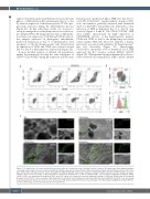
Figure 5. 3D organisation of hemato-endothelial progenitors within the hemanoids using a lentiviral reporter construct. (A) Upper panel: flow cytometry analysis correlating enhanced green fluorescent protein (eGFP) and CD144 expression in embryoid bodies (EB) and hemanoids (day 8 and day 16) generated from the trans- genic human induced pluripotent stem cell line hCD34iPSC16 CBX3.CD144.GFP. Lower panel: analysis of CD34 and CD45 expression in GFP+/CD144+ cells (repre- sentative data of n=3, percentage of gated population is indicated). (B) Expression of GFP in CD144+/CD45– (green; hemato-endothelial progenitors [HEP]), CD144–/CD45+ (red, hematopoietic progenitors [HP]), and CD34– (grey) cells. Upper plot represents individual staining of CD45 and CD144 in day 16 hemanoids (pre-gated on total CD34+ cells, percentage of gated population is indicated). Lower histogram overlay shows GFP expression in HEP, HP and CD34– cells, percentage of gated population is indicated. (C) Fluorescence microscopy of EB and developing hemanoids at day 8 and day 16 of differentiation (brightfield, CD144. GFP fluo- rescence and overlay, scale bar 200 mm and 100 mm respectively).
1360
haematologica | 2021; 106(5)
