
Page 139 - 2021_05-Haematologica-web
P. 139
3D iPSC-differentiation model
ation process. To this end, EB were generated from an established iPSC line (hCD34iPSC16)28 by culture on an orbital shaker for 5 days and subsequently, cultured in media (i) without cytokines, (ii) with IL-3 only and (iii) in standard conditions containing IL-3/M-CSF (Figure 1A). While the absence of any cytokines precluded the produc- tion of hematopoietic cells from hemanoids, supplementa- tion with IL-3 alone resulted in the continuous production of predominantly (>90%) CD45+ blood cells that could be harvested from the supernatant over several weeks, albeit at 2-3-fold lower quantities compared to the standard IL- 3/M-CSF condition (Figure 1B; Online Supplementary Figure S1A). Interestingly, cells produced by the IL-3 only hemanoids were smaller (based on their forward scatter profile/properties in flow cytometry) and showed a more immature myeloid phenotype of CD45+/CD11b+/CD14–/CD163– compared to the predom- inantly CD45+/CD11b+/CD14+/CD163+ and larger mono- cyte/macrophages generated by the standard IL-3/M-CSF culture condition (Figure 1C-D; Online Supplementary Figure S1A). Cells harvested from IL-3 only cultures showed a higher frequency of cells with colony forming potential, as demonstrated by a significantly higher potential to form granulocyte/monocyte colonies (CFU-GM) and erythroid burst forming units (BFU-E) or granulocyte/erythrocyte/monocyte/megakaryocyte (CFU- GEMM) colonies in a methyl-cellulose-based assay (Figure 1E). Of note, the few cells generated without cytokine supplementation did not give rise to any colonies in this assay. Cells from the IL-3 only hemanoids could be further matured into iPSC-derived macrophages or granulocytes in the presence of M- or G-CSF, respectively. iPSC-derived macrophages were characterized by the expression of CD14 and CD163, whereas iPSC-derived granulocytes showed expression of CD66b and CD16 upon final differ- entiation (Figure 1F; Online Supplementary Figure S1B). Taken together, we show that addition of IL-3 only is suf- ficient to induce the hematopoietic program in our
hemanoid differentiation system and enables the genera- tion of iPSC-derived myeloid progenitor cells.
Hematopoietic differentiation in human induced pluripotent stem cell-derived hemanoids recapitulates embryonic hematopoietic development
As a next step, we aimed to further characterize the cel- lular composition of the hemanoids and performed hema- toxilin/eosin (H/E) staining on paraffin embedded sec- tions. Whereas a high proportion of EB resembled neuro- ectodermal structures, after hematopoietic specification, several hemanoids also displayed glandular, vimentin-pos- itive mesodermal and pan-cytokeratin-positive epithelial structures (Figure 2A; Online Supplementary Figure S2A). Moreover, several hemanoids of day 16 of differentiation demonstrated cystic structures with endothelial linings as well as hematopoietic cells stained positive for CD45 (Figure 2A and B). Immunofluorescent staining moreover confirmed the development of all three germlayers in the hemanoids as we detected tubulin positive cells marking neuronal networks, FOXA2 positive endoderm as well as desmin positive mesodermal/muscle cells (Online Supplementary Figure S2B).
In order to further delineate the effect of IL-3 on early hematopoietic specifications, we next investigated early key stages of human embryonic hematopoietic develop- ment in our hemanoid system. We dissociated EB and whole hemanoids cultured with IL-3 at defined differenti- ation stages (days 4, 8 and 12) and analyzed the expression of specific TF associated with hematopoietic specification and the emergence of early hematopoietic cell popula- tions. We found differential regulation of key genes involved in early human hematopoietic development within the first 16 days of differentiation. The primitive streak marker MIXL1 and early mesoderm gene KDR1 were upregulated during EB formation and subsequently progressively downregulated in the hematopoietic specifi- cation phase. Transient expression of SOX17, a key TF
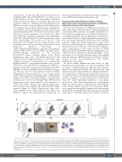
A
C
B
D
Figure 4. Characterization of hematopoietic progenitors within hemanoids. (A and B) Frequency of CD34+CD45+ hematopoietic progenitors (HP) at different days of hematopoietic specification. (A) Representative flow cytometry data (percentage of gated population is indicated) and (B) quantification of n=4, mean±standard error of the mean (SEM). (C) Left: Frequency of colony-forming unites (CFU) of fluorescence-activated cell sorting-purified HP (mean±SEM, n=4, two biological and two technical replicates) and right: bright field microscopy of colony forming units (CFU) in methylcellulose. (D) Representative cytospin staining of CFU from HP derived from the clonogenic assay (scale bar: 20 mm). *P<0.05, ****P<0.0001; statistical significance was assessed using one-way ANOVA with the Dunnetts multiple com- parison test.
haematologica | 2021; 106(5)
1359
