
Page 137 - 2021_05-Haematologica-web
P. 137
3D iPSC-differentiation model
serum (FCS) (Hyclone), 1 mM L-glutamine, 1% NEAA, 1% peni- cillin/streptomycin (all from Invitrogen) and 0,2% b-mercap- toethanol (Sigma-Aldrich). In order to counteract transgene silenc- ing and maintain a high expression of DLL, cells were cultured with 2 mg/mL puromycin (Roth). Cells were passaged twice a week using trypsin. For EHT assay, OP9 DLL1 and DLL4 cells were mixed 50:50 and seeded in 12-well plates (TPP, Switzerland).
Endothelial to hematopoietic transition culture
In order to facilitate endothelial to hematopoietic transition out- side the hemanoids, fluorescence-activated cell sorting (FACS)-
purified HEP (CD34+/CD144+/CD45–) were cultured on over- grown OP9 stromal cells expressing DLL1 and DLL4 in differenti- ation medium I supplemented with 50 ng/mL IL-3 (or as a control in the absence of any cytokines). Cells were analyzed by microscopy or flow cytometry 7-14 days post sorting.
Ethical approval
Generation and use of hiPsc has been approved by the local eth- ical committee of Hannover Medical School (Approval No.: 2127- 2014 and 7870_BO_K_2018). Human CD34 cells were isolated following written informed consent of the donor, which has been
AB
C
D
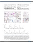
Figure 2. Characterization of hemanoids during hematopioietic specification. (A) Analysis of paraffin sections of hemanoids day 16. A: glandular structure (white arrow), immature mesodermal (black arrow) and neuroectodermal structures (spotted arrow), B: mesodermal structures of varying degrees of differentiation, C: neu- roectodermal formation (CD56 positive [H]), D: smooth muscle actin (ATCA2) positive myoepithelial cells in glandular structures, E: vimentin (VIM) positive mesoder- mal cells, F: pan cytokeratin (CK) positive epithelial structures with varying degrees of positivity, G: PGM1/CD68 mononuclear MO precursors (black arrow) with immature mesoderm and glandular structures (white and spotted arrowheads respectively), H: CD56 positive neuroectodermal formation. Scale bar represents 100 mm). (B) Immunohistochemical analyses of CD45 expression in hemanoids day 16 (bottom panel: arrowheads indicate CD45 positive cells in the magnified area). (C) Analysis of transcription factor expression at defined stages of differentiation (induced pluripotent stem cell [iPSC], embryoid bodies [EB], hemanoids day 4, 8 and 12 and iPSC-derived macrophages [iPSC-MΦ]) by quantitative real time polymerase chain reaction (qRT-PCR) (n=3, mean±standard error of the mean [SEM]). (D) Analysis of surface marker expression at defined stages of differentiation (iPSC, EB, hemanoids day 4, 8, 12 and 16) by flow cytometry (n=3, mean±SEM). *P<0.05, **P<0.01, ***P<0.001, ****P<0.0001 statistical significance was assessed using one-way ANOVA with Dunnetts multiple comparison test.
haematologica | 2021; 106(5)
1357
