
Page 142 - 2021_05-Haematologica-web
P. 142
M. Ackermann et al.
AB
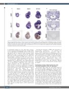
Figure 7. In situ hybridization analyses on embryonic day (E) 9.0, E9.5 and E10.5 wild-type embryos. (A) Whole mount in situ hybridization analysis of IL-3R, Sox17 and Gata2 mRNA in E9.0, E9.5 and E10.5 embryos. IL-3R was expressed in the aorta-gonad-mesonephros (AGM) region (see arrowhead), the tailbud mesenchyme, the limbs, the branchial arches and the somites from E9.0 until E10.5. Sox17 mRNA was expressed in these tissues at all analysed stages. Gata2 transcripts were found in the caudal head mesenchyme from E9.0 until E9.5 and additionally in the otic vesicle, the midbrain and the AGM region at E10.5. (B) RNA in situ hybridiza- tion analysis of transverse sections of the E9.5 embryo.
in endothelial medium for more than three passages, revealed similar endothelial morphology to human umbil- ical cord endothelial cells (HUVEC), and were able to form endothelial tubes in semisolid culture medium (Online Supplementary Figure 3A and B). Besides this endothelial phenotype and functionality, HEP were also analyzed for their potential to undergo the EHT and to give rise to CD34+/CD45+ hematopoietic progenitor cells. Coculture of FACS-purified, CD34+/CD144+/CD45– HEP from hemanoid day 8 on OP9-DLL1 and 4 expressing stroma cells in IL-3 supplemented medium resulted in the forma- tion of characteristic “cobblestone areas” and round- shaped suspension cells after approximately 1 week (Figure 3E). Flow cytometry confirmed the downregula- tion of CD144 as well as the upregulation of CD43 and CD45 in the majority of CD34+ cells and thus the acquired hematopoietic phenotype (Figure 3F; Online Supplementary Figure S3C). CD144–/CD34+/CD45+ hematopoietic stem/progenitor cells from the EHT culture demonstrated high clonogenic potential in a methylcellu- lose assay and gave rise to different colony types with comparable frequency to cord-blood-derived CD34+ cells (Figure 3G; Online Supplementary Figure 3D). iPSC-derived hematopoietic cells isolated from the CFU represented dif- ferent myeloid celltypes such as macrophages, granulo- cytes and erythrocytes (Online Supplementary Figure S3C). In constrast, HEP freshly purified from hemanoids and before EHT did not give rise to hematopoietic colonies when cultured in methyl-cellulose (Online Supplementary Figure S3E).
Following the emergence of HEP in the early phases of hematopoietic specification, we detected first CD34low/CD45+ hematopoietic progenitors (HP) at hemanoid day 4 which increased till hemanoid day 16 to 5.36±1.46 (mean±SD, n=4) (Figure 4A and B). HP were fur- ther characterized by the absence of CD144 and TRA-1- 60, low expression of CD43, and expression of the IL- 3Rα/CD123 (Online Supplementary Figure S3F). Functionally, HP demonstrated clonogenic potential in a methylcellulose-based assay and generated different colony types as well as several myeloid cell types (Figure 4C and D).
Endothelium in human induced pluripotent stem cell-derived hemanoids shows 3D organization
Interestingly, HEP within the developing hemanoids demonstrated a highly organized 3D structure, indicated by a reporter iPSC line (hCD34iPSC),16 which was trans- duced with a lentiviral vector (CBX3.CD144.GFP) express- ing an enhanced green fluorescent protein (eGFP) under the control of a CD144 promoter sub-fragment (modified from).30 In order to prevent epigenetic silencing, a minimal CBX3 ubiquitous chromatin opening element31 was incor- porated into the lentiviral vector. Flow cytometric analysis of EB and hemanoids generated from the reporter line confirmed a strong and specific eGFP expression only in CD34+/CD144+/CD45– cells, as well as a downregulation of eGFP expression in CD34+/CD144–/ CD45+ hematopoi- etic progenitor cells (Figure 5A-B). When analyzing the developing hemanoids derived from the CD144-reporter
1362
haematologica | 2021; 106(5)
