
Page 143 - 2021_05-Haematologica-web
P. 143
3D iPSC-differentiation model
iPSC line by fluorescence microscopy, we noted GFP expression already at the EB stage, which correlated with the emergence of few HEP detected by flow cytometry. Following differentiation, an organized and arborizing endothelial like network was formed within the hemanoids, as seen by fluorescence microscopy on differ- ent days of differentiation (Figure 5C).
Transcriptome analysis recapitulates defined developmental stages
We next subjected FACS purified TRA-1-60+ pluripotent iPSC, TRA-1-60-/KDR+ mesodermal progenitor cells derived from EB, CD34high/CD144+/CD45– HEP and CD34low/CD144–/CD45+ HP derived from the hemanoids to whole transcriptome analysis. Quantitative RT-PCR on
AB
CD
E
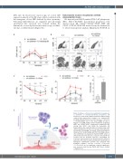
Figure 8. Influence of IL-3 on the endothelial to hematopoiet- ic transition of hemato-endothelial progenitors. (A) Frequency of total CD34+ within hemanoids at different days cultured either without (w/o) cytokines, w/o cytokines+IL-3 blocking antibody (IL-3 block) or in the presence of IL-3 (n=4- 7, mean±standard error of the mean [SEM]). (B and C) Analysis of CD144+/CD45- hemato-endothelial progenitors (HEP) and CD144–/CD45+ hematopoietic progenitors (HP) among total CD34+ cells within the hemanoids. (B) Representative flow cytometry data for hemanoid day (d) 8 and (C) quantification of n=4-7 over 16 days of hematopoietic specification, mean±SEM, percentage of gated population is indicated). (D) Colony forming units (CFU) in methyl-cellulose after endothelial to hematopoietic transition (EHT) culture on OP9 stromal cells in the presence or absence of IL-3 (n=4, two technical and two biological replicates, mean±SEM). (E) Schematic representation of the hemanoid model system as well as the effect of IL-3 on the hematopoietic specification of hemogenic endothelium (scheme was prepared with BioRender software). *P<0.05, **P<0.01, ***P<0.001, ****P<0.0001, ns: not significant; statistical significance was assessed using two-way ANOVA with Tukey’s multiple comparison test.
haematologica | 2021; 106(5)
1363
