
Page 57 - 2021_04-Haematologica-web
P. 57
Combining ibrutinib and checkpoint blockade in CLL
15% decrease in the Nr4a1-GFP MFI at 100 nM and abro- gation of Nr4a1-GFP induction in most of the cells at a concentration of 1 mM (Figure 3A). Similarly, the induction of early activation marker CD69 was significantly reduced at a concentration of 100 nM or higher (Figure 3A). We compared these effects to the more specific BTK inhibitors, CC-292 and ACP-196, which have ITK IC50 val- ues of 24 and 1,000 nM, respectively, compared to that of 4.9 nM of ibrutinib.32 Interestingly, we observed that the decrease in Nr4a1-GFP and CD69 signals was less pro- nounced in CC-292 and was entirely absent upon ACP- 196 treatment (Figure 3A), indicating that these effects are BTK-independent. In line with these results, ibrutinib, but not ACP-196 treatment of mouse splenocytes, inhibited the expression of other activation markers like CD25, CD44 and CD137 on CD8+ T cells in response to αCD3 stimulation (Figure 3B and Online Supplementary Figure S3A-C). In addition, CFSE dilution assays showed that ibrutinib, but not ACP-196, inhibited CD8+ T-cell prolifer- ation (Online Supplementary Figure S3D). Similar to murine
splenocytes, treatment of human PMBC with ibrutinib resulted in a dose-dependent decrease in proliferation of CD8+ T cells in response to αCD3 stimulation (Figure 3C). Collectively, these data suggest that ibrutinib can modu- late CD8+ T-cell response to TCR stimulation in a BTK- independentmanner.
+
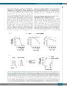
Strong co-stimulatory signals can rescue CD8 T cells
from inhibitory effects of ibrutinib in vitro
We next evaluated potential approaches to unleash
CD8+ T cells from ibrutinib’s inhibitory effects. Previous work has shown that CD28 signaling is intact in the absence of ITK.33 Thus, we hypothesized that strong CD28 co-stimulation can hijack ibrutinib inhibitory effects on CD8+ T cells. Therefore, we evaluated the effect of the drug on murine CD8+ T cells stimulated with αCD3 alone or αCD3 plus αCD28. The addition of αCD28 anti- body had no impact on the drop of Nr4a1-GFP signal after ibrutinib treatment (Figure 4A), which is in line with pre- vious work demonstrating that Nr4a1 induction by TCR
A
BC
Figure 4. Strong co-stimulatory signals can rescue CD8+ T cells from inhibitory effects of ibrutinib in vitro. (A) (Left) Splenocytes from Nr4a1-GFP transgenic mice (n=3) were pretreated with different concentrations (Conc.) of ibrutinib or dimethyl sulfoxide (DMSO) for 30 minutes (min), and then stimulated with either αCD3 or αCD3 plus αCD28 antibodies for 6 hours (h). Nr4a1-GFP, expression was analyzed by flow cytometry in viable, 7-aminoactinomycin D (7-AAD)-negative, single CD8+ T cells. (Middle and right panels) Splenocytes from C57BL/6 mice (n=4) were pretreated with different concentrations of ibrutinib or DMSO for 30 min and then stim- ulated with either αCD3 or αCD3 plus αCD28 antibodies. CD25, and CD44 expression were analyzed by flow cytometry in CD8+ T cells after 24 h. (B) Splenocytes from C57BL/6 mice (n=4) were labeled with carboxyfluorescein succinimidyl ester (CFSE), pretreated with different concentrations of ibrutinib or DMSO for 30 min and then stimulated with αCD3 or αCD3 plus αCD28 antibodies. Proliferation as measured by CFSE dilution after 48 h was analyzed by flow cytometry in viable, 7- AAD-negative, single CD8+ T cells. (C) Peripheral blood mononuclear cells (PBMC) from healthy donors (n=3) were labeled with CFSE, pretreated with different con- centrations of ibrutinib or DMSO for 30 min and then stimulated with αCD3 or αCD3 plus αCD28 antibodies. Proliferation as measured by CFSE dilution after 72 h was analyzed by flow cytometry in viable, 7-AAD-negative, single CD8+ T cells. Graphs show means±standard error of mean. MFI: median fluorescence intensity.
haematologica | 2021; 106(4)
973
