
Page 55 - 2021_04-Haematologica-web
P. 55
Combining ibrutinib and checkpoint blockade in CLL
treated mice in comparison to untransplanted WT con- trols (Figure 1D and E). Interestingly, the percentage and absolute numbers of the effector population were lower in ibrutinib-treated mice (Figure 1D and E). This was accom- panied by decreased expression of activation markers like CD69 and CD137 on CD8+ T cells (Figure 1F), and decreased expression of transcription factors that regulate effector T-cell differentiation, such as T-box transcription factor T-bet and Eomesodermin (Eomes) (Figure 1G).30 Furthermore, CD8+ T-cell proliferation was significantly lower in ibrutinib- compared to vehicle-treated TCL1 mice, reaching levels similar to non-tumor control mice (Figure 1H). The drop in the effector population, activa- tion markers and proliferation of CD8+ T cells was also observed when treatment started at later stages of disease when all mice had more than 5,000 CLL cells/mL of blood, the leukemic threshold in CLL patients (Online Supplementary Figure S1A).
We next examined whether the observed changes in CD8+ T cells are caused by a direct impact of ibrutinib on these cells or rather reflect a secondary normalization of the T-cell compartment due to the decrease in tumor load. Thus, we analyzed CD8+ T cells in vehicle- and ibrutinib- treated mice having a similar disease load, as measured by
CLL-cell infiltration in spleen and liver. While the vehicle- treated mice showed signs of overt leukemia with severe hepato-splenomegaly after 3 weeks of starting the treat- ment, ibrutinib successfully suppressed CLL development during that time. Nonetheless, mice continuously receiv- ing ibrutinib succumbed to full-blown leukemia at week 4 post treatment exhibiting similar disease load to vehicle group at week 3 (Online Supplementary Figure S1B). Analysis of the spleen CD8+ T-cell compartment of end- stage vehicle or ibrutinib-treated mice with comparable tumor load revealed a decrease in effector T-cell percent- age and numbers, accompanied by a significant drop in activation marker expression and proliferation in the latter group (Online Supplementary Figure S1C-E). This suggests that the observed differences in CD8+ T cells in ibrutinib- versus vehicle-treated mice were not secondary to changes in tumor load.
Ibrutinib modulates CD8+ T-cell function in the TCL1 adoptive transfer model
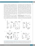
We next examined the impact of ibrutinib treatment on the expression of inhibitory receptors like PD-1, CD244 and Lag3 on CD8+ T cells. We observed a substantial drop in the expression of these markers in the ibrutinib cohort
AB
CD
Figure 2. Ibrutinib modulates CD8+ T-cell function in the TCL1 adoptive transfer model. C57BL/6 mice were transplanted with splenocytes from leukemic TCL1 mice, and after 2 weeks assigned to treatment with ibrutinib (0.16 mg/mL) or vehicle control in drinking water. Mice were sacrificed after 2 weeks of treatment. (A) Flow cytometric analysis of PD-1, CD244, and Lag3 expression on CD8+ T cells from vehicle- or ibrutinib-treated mice (n=4). (B-D) Cytotoxic function of CD8+ T cells was assessed by flow cytometric analysis of (B) degranulation capacity, as measured by CD107a expression on the cell surface, (C) GzmA and GzmB expression or (D) IFNγ production (n=4). Graphs show means±standard error of mean. *P<0.05, **P<0.01, ***P<0.001. nMFI: normalized median fluorescence intensity.
haematologica | 2021; 106(4)
971
