
Page 56 - 2021_04-Haematologica-web
P. 56
B.S. Hanna et al.
(Figure 2A). As T-cell inhibitory receptors are mainly induced upon activation of tumor-reactive T cells,31 we evaluated whether the drop in their expression reflects an improvement in T-cell function or is rather a consequence of the drop in T-cell activation in ibrutinib-treated mice (Figure 1F). Thus, we analyzed the functional capacities of CD8+ T cells using intracellular flow cytometric staining fol- lowing mitogen PMA/ionomycin stimulation. CD8+ T cells from ibrutinib-treated mice exhibited lower degranulation potential, as measured by CD107a presentation on the cell surface (Figure 2B). Furthermore, granzyme A (GzmA) and GzmB expression in CD8+ T cells significantly dropped in these mice (Figure 2C). Moreover, production of IFNγ was significantly lower in CD8+ T cells from ibrutinib-treated mice (Figure 2D), collectively indicating poor cytotoxic and effector function of these cells. The changes in cytokine production and cytolytic abilities were primarily attributed to the decrease in the effector CD8+ fraction after ibrutinib treatment. Nonetheless, degranulation capacity, GzmA and GzmB levels were also decreased when focusing the analy- sis on the minor effector or PD-1+ population in ibrutinib- treated mice (Online Supplementary Figure S2A-F). In summa- ry, these data indicate that in vivo ibrutinib treatment alters
activation, effector differentiation and function of CD8+ T cells in the TCL1 AT model.
Ibrutinib modulates T-cell receptor signaling in CD8+ T cells in vitro
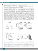
We then investigated the mechanisms that control the above-mentioned changes in T-cell function. While ibruti- nib can indirectly impact T cells through BTK inhibition in CLL cells or other antigen-presenting cells in the microen- vironment, the drug can potentially affect T-cell function in a direct manner through its off-target effects on ITK. To examine these possibilities, we tested the effect of differ- ent BTK inhibitors on TCR signaling using a reporter mouse line for Nr4a1, an immediate target of TCR activa- tion.24 Splenocytes from Nr4a1-GFP mice were pre-treated with ibrutinib or dimethyl sulfoxide (DMSO) as control and then stimulated with αCD3 for 6 hours (h). We prima- rily used ibrutinib in a dose range of 100-1,000 nM, which results in ITK occupancy of up to 50% corresponding to the level observed in vivo in ibrutinib-treated CLL patients.15 αCD3 stimulation resulted in a rapid increase of the Nr4a1-GFP signal in CD8+ T cells in DMSO controls (Figure 3A). Interestingly, ibrutinib treatment resulted in a
A
BC
Figure 3. Ibrutinib modulates T-cell receptor signaling in CD8+ T cells in vitro. (A) Splenocytes from Nr4a1-GFP transgenic mice (n=3) were pretreated with different concentrations of ibrutinib, ACP-196, CC-292 or DMSO for 30 minutes (min), and then stimulated with αCD3 antibody for 6 hours (h). Nr4a1-GFP (left panels) and CD69 (right panels) expression was analyzed by flow cytometry in viable, 7-aminoactinomycin D (7-AAD)-negative, single CD8+ T cells. (Right) Relative percentages of Nr4a1-GFP- or CD69-positive cells. FMO: fluorescence minus 1. (B) Splenocytes from C57BL/6 mice (n=3) were pretreated with different concentrations of ibru- tinib or DMSO for 30 min and then stimulated with αCD3 antibody. CD25, CD44 and CD137 expression was analyzed by flow cytometry in viable, 7-AAD-negative, single CD8+ T cells after 24 h. (C) Peripheral blood mononuclear cells (PBMC) from healthy donors (n=5) were labeled with 5 mM carboxyfluorescein succinimidyl ester (CFSE), pretreated with different concentrations of ibrutinib or DMSO for 30 min and then stimulated with αCD3 antibody. Proliferation as measured by CFSE dilution after 72 h was analyzed by flow cytometry in viable, 7-AAD-negative, single CD8+ T cells. Graphs show means±standard error of mean. *P<0.05, **P<0.01, ***P<0.001; MFI: median fluorescence intensity.
972
haematologica | 2021; 106(4)
