
Page 58 - 2021_04-Haematologica-web
P. 58
B.S. Hanna et al.
ABC
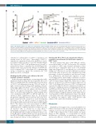
Figure 5. Blocking PD-1/PD-L1 axis enhances the anti-leukemic activity of ibrutinib. C57BL/6 mice were transplanted with splenocytes from leukemic TCL1 mice, and after 2 weeks assigned to treatment with isotype antibody plus vehicle (control), αPD-1, αPD-L1, ibrutinib, αPD-1 + ibrutinib, or αPD-L1 + ibrutinib. Mice were sacrificed after 4 weeks of treatment (n=6). (A) Absolute numbers of CD5+CD19+ chronic lymphocytic leukemia (CLL) cells in peripheral blood over time analyzed by flow cytometry, and (B) spleen weight of different treatment arms at the end-point (n=6). (C) Tumor load, defined as percentage of CD5+CD19+ CLL cells out of CD45+ cells in bone marrow (BM) (n=4-6). Graphs show means±standard error of mean. *P<0.05, **P<0.01, ***P<0.001.
stimulation is independent of CD28 co-stimulation and remains intact in cd28-/- mice.24 Interestingly, CD28 co- stimulation completely abolished the inhibition of activa- tion marker expression that is caused by ibrutinib (Figure 4A). Moreover, CD8+ T-cell proliferation was rescued to normal levels when co-stimulated by αCD28 (Figure 4B). Similar results were observed by addition of αCD28 anti- body to human CD8+ T cells (Figure 4C), confirming that strong co-stimulatory signals can circumvent the TCR inhibitory effects of ibrutinib.
Blocking the PD-1/PD-L1 axis enhances the anti- leukemic activity of ibrutinib
In the subsequent experiments, we investigated possible immunomodulatory drugs that can recapitulate the in vitro effects of αCD28 antibodies and thereby reverse the block of CD8+ T-cell differentiation in the TCL1 AT model. In light of recent results showing CD28 signaling as the pri- mary target of PD-1 blockade,34,35 we reasoned that block- ing the PD-1/PD-L1 axis can enhance anti-tumoral T-cell activity and improve therapeutic efficacy of ibrutinib. Thus, we treated CLL-bearing mice with ibrutinib alone or in combination with αPD-1 or αPD-L1 antibodies. As shown in Online Supplementary Figure S4, all treatment groups had comparable tumor load at the start of the treat- ment. Consistent with our previous work, blocking the PD-1/PD-L1 axis by αPD-L1 resulted in delayed CLL development in PB, spleen and bone marrow (BM) (Figure 5A-C). The effects were more pronounced for αPD-L1 compared to αPD-1 treatment, which is likely due to the additional effects of αPD-L1 antibodies on tumor-associat- ed myeloid cells, as shown by us and others.9,36 Interestingly, combination of ibrutinib with PD-1 or PD- L1 blocking antibodies resulted in enhanced disease con- trol in PB, spleen and BM, which was most pronounced in the αPD-L1/ibrutinib combination (Figure 5A-C), confirm- ing a therapeutic efficacy of this combination.
Blocking the PD-1/PD-L1 axis expands the effector population and enhances the functional capacity of CD8+ T cells
We then examined the effect of PD-1/PD-L1 combina- tion with ibrutinib on CD8+ T-cell activity. We restricted our analysis to αPD-1 plus ibrutinib treatment as these mice had more comparable tumor load to the ibrutinib single arm (Figure 5A-C), and thereby we could exclude changes in T cells that are caused by differences in disease load. In line with our previous data, ibrutinib reduced the percentage of effector population, and decreased activa- tion, proliferation and functional capacities of CD8+ T cells (Figure 6A-D). Interestingly, combination of ibruti- nib with αPD-1 resulted in a significant increase in effector CD8+ T-cell percentages (Figure 6A), which was accompa- nied by an increase in activation marker expression on CD8+ T cells (Figure 6B). Moreover, CD8+ T-cell functional capacity was improved by the combination treatment, as shown by a significant increase in degranulation capacity and IFNγ production (Figure 6C and D). The increase in degranulation capacity and IFNγ production was also observed when focusing the analysis on the CD8+ effector T-cell population (Online Supplementary Figure S5A and B). Collectively, these data show that blocking the PD-1/PD- L1 pathway can improve CD8+ T-cell function and enhance CLL control after ibrutinib treatment.
Discussion
Ibrutinib has been approved as a successful inhibitor of BTK for treatment of CLL and other B-cell lymphomas. In addition, it was also the first clinically available inhibitor of ITK.15 ITK is expressed in T cells in which, as a major player in TCR signaling, it is responsible for calcium mobilization, cytoskeleton reorganization, synapse for- mation and adhesion.37 Deletion or inhibition of ITK in
974
haematologica | 2021; 106(4)
