
Page 59 - 2021_04-Haematologica-web
P. 59
Combining ibrutinib and checkpoint blockade in CLL
A
BCD
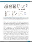
Figure 6. Blocking the PD-1/PD-L1 axis expands the effector population and enhances the functional capacity of CD8+ T cells. C57BL/6 mice were transplanted with splenocytes from leukemic TCL1 mice, and after 2 weeks assigned to treatment with isotype antibody plus vehicle (control), αPD-1, ibrutinib, or αPD-1 + ibrutinib. Mice were sacrificed after 4 weeks of treatment. (A) Flow cytometric analysis of splenic CD3+CD8+ T cells from different treatment arms (n=4). Cell subsets were defined as naïve (CD127hiCD44low), memory (Mem: CD127hiCD44hi), and effector (Eff: CD127lowCD44int-hi) cells. (B) Flow cytometric analysis of CD69 expression. (C and D) Cytotoxic function of CD8+ T cells was assessed by flow cytometric analysis of (C) degranulation capacity, as measured by CD107a expression on the cell sur- face, and (D) IFNγ production (n=4). Graphs show means±standard error of mean. *P<0.05, ***P<0.001, ns: not significant.
CD8+ T cells results in their decreased expansion, delayed expression of cytolytic effector molecules, and defective degranulation upon TCR activation.38 This leads to a glob- al defect in the cytolysis of pathogens, and as a conse- quence, to reduced viral clearance. In line with this, dra- matic in vivo immunomodulation by ibrutinib has led to its approval for the treatment of refractory cGvHD,20 but the mechanisms underlying this clinical efficacy are poorly understood.
So far, modulating effects of ibrutinib on the differenti- ation of CD4+ T cells, like Th1, Th2, Th17 and Treg cells, have been described.15,19,22 In the current study, we observed a negative impact of ibrutinib on CD8+ T-cell proliferation and function in the TCL1 AT model, which reduces their phenotype to a level of tumor naïve control mice. As our in vitro data confirmed CD8+ T-cell inhibition by ibrutinib, but not the specific BTK inhibitor ACP-196, this effect is BTK-independent and most likely mediated by inhibition of ITK downstream of the TCR. Along this line, CC-292, an inhibitor with a higher selectivity for BTK, had a lower impact on T-cell activity in vitro, and has been previously shown not to impair T-cell function in the TCL1 AT model.39 But as these drugs might have different efficacies in patients, caution must be exercised when extrapolating clinical effects from these pre-clinical obser- vations. Our observations in the TCL1 AT model seem to be in contrast to published data describing an increase of CD8+ T-cell numbers and their reduced expression of inhibitory receptors, like PD-1, in CLL patients after 8-20 weeks of treatment with ibrutinib.19 The results of this study suggest that this may be due to diminished activa- tion-induced cell death of T cells through ITK inhibition. As our data demonstrate that inhibition of ITK by ibruti-
nib decreases TCR signaling and thereby activation of T cells, a consequence of that is not only reduced function- al properties, as we show, but also diminished activation- induced cell death, confirming the observations of Long et al. in CLL patients.19 A further study documented that ibrutinib decreases CD8 T-cell proliferation and activation in patients 8 weeks after treatment start.22 However, these results have been largely viewed as a sign of favorable immune normalization under ibrutinib treatment, rather than a direct side effect of the drug. In light of our in vitro and in vivo results, it is more likely that these findings in CLL patients are due to the off-target effects of ibrutinib on ITK. In a further study that investigated T cells in CLL patients before and after 1 year of treatment with ibruti- nib, disease-associated, elevated T-cell numbers and T-cell-related cytokine levels in PB had normalized, and T-cell repertoire diversity had increased significantly in treated patients.40 Considering the broad effects of BTK blockade on functional properties of many immune cell types, these changes in the T-cell compartment might be due to ibrutinib’s ability to rewire the carefully construct- ed, supportive microenvironmental network in CLL, which presumably contributes to the therapeutic success of this drug.41 But as ibrutinib, first-of-all, considerably redistributes malignant B cells from lymph nodes to blood, which ultimately results in a reduction of tumor burden, this suggests that normalization of the T-cell compartment under ibrutinib treatment is secondary to the effect of the drug on CLL-cell numbers and their localization. This is supported by our previous results as well as published data of others showing that T-cell expansion and exhaus- tion positively correlates with tumor load and predomi- nantly occurs in secondary lymphoid organs.7-9 Along this
haematologica | 2021; 106(4)
975
