
Page 154 - 2021_02-Haematologica-web
P. 154
A. Korporaal et al.
AB
CD
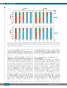
Figure 3. Developmental expression patterns of human β-like globins in control and Klf1wt/Nan embryos. Expression of he, hγ and hβ was determined by reverse tran- scriptase quantitative PCR. Data are displayed as fraction of total hβ-like globin (he+hγ+hβ) expression. Embryonic day (E) and number of embryos (N) are indicated. *P<0.05; error bars indicate standard deviations.
of nucleated cells remained significantly higher in the Klf1wt/Nan samples in all E14.5 litters.
Using a CASY cell counter, we determined the cell size distributions in E14.5 blood samples. In E14.5 control sam- ples, the two peaks representing primitive (large) and definitive (small) cells are clearly separated (Figure 4C). In E14.5 Klf1wt/Nan samples, these two peaks are not clearly separated. The apparent continuum of cell sizes is in agreement with the rampant anisocytosis observed in the cytospins of E14.5 Klf1wt/Nan blood (Figure 4A). Finally, we sought to use flow cytometry as an orthogonal approach to determine the contribution of primitive erythrocytes to the circulation. In an attempt to better distinguish primi- tive from definitive erythrocytes, we performed flow cytometry using CD71 (transferrin receptor), Ter119, and CD9 (Tetraspanin) which is a marker for primitive ery- throcytes.35 Compared to the controls, expression of CD71 was slightly increased on Klf1wt/Nan E10.5 primitive cells (Figure 4D). This might indicate a delay in matura- tion, similar to what has been proposed for Klf1wt/ko reticu- locytes.36 Expression of Ter119 was virtually absent in E10.5 Klf1wt/Nan erythrocytes, while CD9 expression was strongly reduced (Figure 4D). At E14.5, CD9 was unable to distinguish primitive from definitive erythrocytes in Klf1wt/Nan blood, in contrast to E14.5 Klf1wt/Nan blood in which a distinct fraction of CD9+ primitive cells was observed (Figure 4D, arrow). Collectively, we conclude that the con- tribution of primitive erythrocytes to the circulation of Klf1wt/Nan embryos cannot be determined by flow cytome-
try using CD71, Ter119 and CD9 as markers. Despite these technical limitations, the analysis of blood samples is consistent with the notion that, compared to control embryos, the contribution of primitive erythrocytes to the pool of circulating erythrocytes in Klf1wt/Nan embryos is extended during development.
Klf1wt/Nan erythroblasts display impaired proliferation and differentiation
The delays in hemoglobin switching and appearance of definitive erythrocytes in the circulation of Klf1wt/Nan embryos suggest that the production of definitive cells in the fetal liver might be affected by impaired proliferation or differentiation. Since KLF1 is critically involved in regu- lation of the erythroid cell cycle,11,37,38 central to both these processes, we cultured primary cells derived from E12.5 fetal livers to assess the proliferative capacity of Klf1wt/Nan erythroblasts. RT-qPCR analysis at day 6 of culture showed deregulated expression of the embryonic/fetal globin genes in the Klf1wt/Nan cells compared to the controls, demonstrating that this aspect of the phenotype is main- tained in the culture system (Online Supplementary Table S1). We have previously shown that Klf1ko/ko E12.5 fetal liver cells expand well when grown under selfrenewal conditions.9 In contrast, Klf1wt/Nan erythroblasts from E12.5 fetal liver expanded very poorly under these growth con- ditions (Figure 5A). Up to day 3-4 of culture, Klf1wt/Nan fetal liver cells expanded similar to those derived from control embryos. After day 4, expansion of the Klf1wt/Nan cultures
468
haematologica | 2021; 106(2)
