
Page 153 - 2021_02-Haematologica-web
P. 153
Hemoglobin switching in Klf1wt/Nan mice
AB
CD
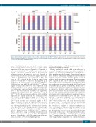
Figure 2. Developmental expression patterns of mouse α-like globins in control and Klf1wt/Nan embryos. Expression of mz and mα was determined by reverse tran- scriptase quantitative PCR. Data are displayed as fraction of total mα-like globin (mz+mα) expression. Embryonic day (E) and number of embryos (N) are indicated. *P<0.05; error bars indicate standard deviations.
Delayed appearance of definitive erythrocytes in the circulation of Klf1wt/Nan embryos
Having established that the shift from embryonic to fetal and adult globin expression is delayed in Klf1wt/Nan embryos, we investigated whether this could be due to a delay in embryonic development. The erythroid compart- ment changes dynamically during mouse development,33 and any alterations in this dynamic change would be reflected in globin expression patterns. Primitive erythro- cytes originate in the yolk sac and are the sole erythro- cytes in the circulation until E12.5, when the first defini- tive erythrocytes are released from the fetal liver. As fetal liver erythropoiesis gathers momentum, the majority of cells in the circulation are definitive erythrocytes by E14.5. In contrast to primitive erythrocytes, definitive erythro- cytes enucleate before they are released in the circulation. We used this characteristic to determine the contribution of primitive cells to the circulation by making cytospins of peripheral blood collected from E14.5 and E16.5 control and Klf1wt/Nan embryos. Compared to the controls, nucleat- ed erythrocytes were more abundant in cytospins of Klf1wt/Nan blood. They were still easily detected in E16.5 cytospins of Klf1wt/Nan blood, while such cells were virtually absent in control samples (Figure 4A). In order to assess the switch from primitive to definitive erythropoiesis, E14.5 cytospins were split into early, mid and late of litter harvest (Figure 4B). Consistent with the previous results, we observed that the fraction of nucleated cells declined very rapidly at this stage of development, in the controls from ~0.34 at early E14.5 to ~0.01 at late E14.5, and in the Klf1wt/Nan samples from ~0.52 at early E14.5 to ~0.13 at late E14.5. Importantly, compared to the controls the fraction
genes. The E11.5 yolk sac and fetal liver of control embryos expressed very similar ratios of he and hγ, while expression of hβ was very low (Figure 3A). Compared to the controls, E11.5 Klf1wt/Nan yolk sac and fetal liver dis- played a small but significant shift to he expression. Relatively increased he expression was also observed in Klf1wt/Nan E12.5 yolk sac and fetal liver (Figure 3B and D).
Next to the difference in he expression at E12.5, hβ made up ~75% of total hβ-like globins in control com- pared to ~43% in Klf1wt/Nan fetal liver. This apparent lag in switching to hβ expression was also observed in Klf1wt/Nan E13.5 fetal liver, and in E13.5 and E14.5 Klf1wt/Nan yolk sacs (Figure 3 B and D). In control yolk sacs, expression of hβ increased rapidly to ~35% at E13.5, with hβ remaining the most abundantly expressed hβ-like globin accounting for ~50% of the total output of the HBB locus (Figure 3A). Compared to control yolk sacs, expression of hγ in Klf1wt/Nan E13.5 yolk sacs was even higher at ~60%, with hβ expression also rapidly increasing but reaching a lower level of ~25% of hβ-like globins (Figure 3B). At E14.5, the hγ:hβ ratio shifted to 4:96 in control yolk sacs, while in Klf1wt/Nan yolk sacs this ratio remained higher at 28:72 (Figure 3A and B). At E16.5, hβ expression accounted for >97% of total hβ-like globin in all yolk sacs and fetal li- vers, showing that hemoglobin switching had quantita- tively proceeded to the adult profile in both genotypes (Figure 3). We conclude that expression of he is main- tained at higher levels in E11.5-E12.5 Klf1wt/Nan embryos. This is followed by a lag in switching to hβ expression, which favors expression of hγ, in E13.5-E14.5 Klf1wt/Nan embryos. Nevertheless, at E16.5 expression of he and hγ has receded to <3% of total hβ-like globins.
haematologica | 2021; 106(2)
467
