
Page 152 - 2021_02-Haematologica-web
P. 152
A. Korporaal et al.
AB
CD
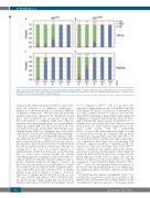
Figure 1. Developmental expression patterns of mouse β-like globins in control and Klf1wt/Nan embryos. Expression of mey, mβh1 and mβ was determined by reverse transcriptase quantitatice PCR. Data are displayed as fraction of total mβ-like globin (mey+mβh1+mβ) expression. Embryonic day (E) and number of embryos (N) are indicated. *P<0.05; error bars indicate standard deviations.
culation in the embryo properly after E8.5 as large nucle- ated cells referred to as primitive erythrocytes.33 Expression of embryonic globins is a distinctive hallmark of primitive erythrocytes. The first intra-embryonically derived erythrocytes appear in the circulation around E12.5. These enucleated cells are generated in the fetal liver and referred to as definitive erythrocytes. They are characterized by predominant expression of adult globins; unlike the human HBB locus the mouse Hbb locus does not harbor fetal β-like globin genes. Nevertheless, mice carrying human HBB locus transgenes have been exten- sively used to study fetal-to-adult hemoglobin switching.34
In order to analyze the developmental dynamics of glo- bin expression in Klf1wt/Nan embryos, we determined the globin expression profiles in RNA isolated from yolk sacs and fetal livers harvested between E11.5 and E16.5. Both the yolk sac and the fetal liver contain tissue cells plus cir- culating blood cells. First, we assessed expression of mβ-like globins. At E11.5, both the yolk sac and the fetal liver of control mice contained mainly mee globin, reflecting the presence of primitive erythrocytes in the cir- culation and the fact that the fetal liver only just starts to produce definitive erythroid cells (Figure 1 A and C). In E12.5 fetal liver the production of large numbers of defin- itive erythroid cells was reflected in the dominant expres- sion of mβ, while meγ still constituted >90% of mβ-like globins in the yolk sac (Figure 1 A and C). At E13.5, expression of mβ-like globins detected in yolk sac was 70% mee and 30% mβ, whereas >95% of fetal liver mβ- like globin was mβ. Finally, mainly mβ was detected in fetal liver and yolk sac from E14.5 onward (Figure 1 A and
C). In comparison, Klf1wt/Nan yolk sac and fetal liver expressed a larger fraction of mβ at both E12.5 and day E13.5 (Figure 1 B and D). The increase of mβ expression over time is delayed in Klf1wt/Nan yolk sac and fetal liver (Figure 1B-D), indicating a delayed shift in the expression of primitive to definitive mβ-like globins. Next, we inves- tigated whether this delay is specific for the Hbb locus, or whether it also occurs in the Hba locus. Yolk sacs and fetal livers from E11.5 control embryos expressed mz as the major α-like globin, with mα contributing 25-30% to total α-like globin expression (Figure 2 A and C). At E12.5 mα became the dominant α-like globin in fetal liver, mz was gradually replaced in the yolk sac by mα at E12.5 and E13.5, with the major shift to mα expres- sion completed by E14.5 (Figure 2A), corresponding with fetal liver output in circulation. A different pattern was observed in yolk sacs and fetal livers from Klf1wt/Nan embryos. Compared to the controls, at E11.5 the contribu- tion of mz was increased at the expense of mα (Figure 2B and D). At E12.5 and E13.5 there was no increase in mα globin expression in the yolk sac (Figure 2B), and the increase in expression of mα in fetal livers was reduced compared to control fetal livers (Figure 2B and D). Thus, Klf1wt/Nan affects the developmental expression patterns of the mouse α-like and β-like globins in a very similar man- ner, displaying a delayed switch to expression of the adult genes.
As CDA-IV patients display very high HbF levels,20-22 we extended the observations on the mouse globins to the human β-like globin genes thus adding analysis of the developmental expression patterns of fetal-stage globin
466
haematologica | 2021; 106(2)
