
Page 164 - 2019_03-Haematologica-web
P. 164
N. Casagrande et al.
44%) and a median CD68 expression level of 17% positive pixels (range, 0%-58%) (Figure 6A). No difference in CCL5 expression was found in EBV-positive versus EBV-negative HL samples (data not shown). On the contrary, a significant correlation between CCL5 and CD68 levels was found (Spearman r=0.251, P=0.0487) (Figure 6B). Patients were then divided into three arbitrary categories of CCL5 expression: low (0%-10%), medium (11%-20%) and high
(21%-100%). These groups had 24, 22, and 16 patients, respective- ly (data missing for 3 patients). A Kaplan-Meier plot showed that the group of patients with high CCL5 expression had significantly worse survival (P=0.0072) than patients with low or medium expression (Figure 6C). The hazard ratio for progression-free sur- vival of low/medium CCL5 expression to that of high expression was 0.015 (P=0.0016; 95% CI, 0.0011-0.2064).
A
B
CD
E
F
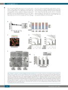
Figure 4. Maraviroc slows tumor cell growth and inhibits heterospheroid formation. (A) cHL cells (2x10⁵ cells/ml) were cultured with increasing concentrations of maraviroc for 72 h, and viable cells were counted by trypan blue dye exclusion. Results are means and SD of three replicate wells from three independent experi- ments. (B) Percentages of cHL cells in the various cell cycle phases after a 24 h treatment with maraviroc (100 mM). Results are means and SD of at least three experiments. (C) Representative image of 3D heterospheroids generated by plating HDLM-2 cells (stained green with CFSE), MSCs (red with fluorescent DiI), and monocytes (blue with DiD) under non-adherent conditions (poly-HEMA-coated wells). (D) L-1236 or HDLM-2 cells, HL-MSCs and monocytes were cultured in RPMI 1640 medium plus 1% FCS, alone or in combination under non-adherent conditions. After 4 days, conditioned medium was collected for CCL5 ELISAs; three different experiments were evaluated. (E) Heterospheroids generated by co-cultivation of cHL cells (L-1236, HDLM-2, or L-540 cells) with HL-MSCs and monocytes under non- adherent conditions in the presence or absence of maraviroc (100 mM) and photographed after 24 h using an inverted microscope (Eclipse TS/100, Nikon). (F) Heterospheroids (cHL + HL-MSCs + monocytes) were cultured with and without maraviroc. After 48 h, viable cells were evaluated using PrestoBlue Cell Viability Reagent. Relative fluorescence units (RFU). Values are means and SD of three experiments. cHL: classical Hodgkin lymphoma; (HL)-MSCs: Hodgkin lymphoma; MSC: mesenchymal stromal cells; 3D: three dimensional.
570
haematologica | 2019; 104(3)
