
Page 162 - 2019_03-Haematologica-web
P. 162
N. Casagrande et al.
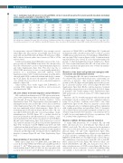
Table 1. Combination index (CI) values for L-1236 and HDLM-2 cell lines treated with maraviroc (first column) and with doxorubicin, brentuximab vedotin, cisplatin, gemcitabine or vinorelbine for 72 h.
MVC Doxo CI BV CI CDDP CI GCB CI VRB CI
L-1236
HDLM-2
(mM) (ng/mL) 25 6.0
50 12.5
100 *25.0
25 3.1
50 6.2
100 *12.5
(mg/mL) (μM) (nM) 0.397 3.8 0.253 0.13 0.606 0.15
0.594 7.5 0.357 0.25 0.955 0.30
0.628 *15.0 0.495 *0.50 1.152 *0.60
0.512 75 0.373 0.31 0.571 0.80
0.526 150 0.478 0.63 0.706 1.60
0.753 *300 0.655 *1.25 1.169 *3.20
0.662 0.912 1.302 0.699 1.054 1.173
(ng/mL)
0.13 0.849
0.25 0.946
*0.50 1.051
0.25 0.818
0.50 0.887
*1.00 1.045
Cell viability was determined by trypan blue dye exclusion. Combination indexes were calculated using CalcuSyn software. *Indicates the IC50 for each drug. (IC50) Concentration required for 50% in vitro inhibition of growth. MVC: maraviroc; Doxo: doxorubicin; BV: brentuximab vedotin; CDDP: cisplatin; GCB: gemcitabine;VRB: vinorelbine
becoming tumor educated (E-BM-MSCs), they strongly secreted CCL5 (Figure 1E). This response was partially reduced by treat- ment with an anti-TNFα antibody (Figure 1F). Education of BM- MSCs did not, however, induce the secretion of CCL3 or CCL4 (data not shown).
Conditioned medium from E-BM-MSCs increased the clono- genic growth of L-1236, HDLM-2 and L-540 cells, and this effect was reduced by maraviroc in a dose-dependent manner (Figure 1G and Online Supplementary Figure S2A). This effect was due to CCL5-CCR5 interactions, because it was partially inhibited by inclusion of a neutralizing anti-CCL5 antibody (Online Supplementary Figure S2B). Conditioned medium from BM-MSCs educated with L-1236-conditioned medium (and thereby contain- ing CCL5; Figure 1E) increased the migration of CD14+ mono- cytes, and this effect was reduced in a dose-dependent manner by maraviroc (Figure 1H).
Taken together, these results suggest that E-BM-MSCs, by secreting CCL5, stimulate tumor growth as well as monocyte recruitment in the TME.
cHL cells induce monocyte migration and proliferation
cHL-conditioned medium increased CCR5 expression in mono- cytes (Online Supplementary Figure S3A) and enhanced their migra- tion through fibronectin-coated Boyden chambers (Online Supplementary Figure S3B). Representative photomicrographs of transmigrated monocytes (red Fast-Dil colored cells) are shown in Online Supplementary Figure S3C. This enhanced monocyte migra- tion was significantly reduced when maraviroc (Figure 2A) or a neutralizing anti-CCL5 antibody (Figure 2B) was added. cHL cells, especially L-1236 and L-428 cells, secreted macrophage colony- stimulating factor (M-CSF) (Figure 2C), a cytokine involved in monocyte proliferation and differentiation. In accordance, condi- tioned medium from cHL cells increased monocyte growth (Figure 2D and Online Supplementary Figure S3D). E-monocytes secreted CCL3 and low amounts of CCL4 and CCL5 (Online Supplementary Figure S3E). Treatment of L-1236, HDLM-2 and L-540 cells with conditioned medium from E-monocytes increased the number of viable tumor cells (Online Supplementary Figure S3F) and stimulated clonogenic growth (Figure 2E), but this growth was inhibited by maraviroc treatment (Figure 2E and Online Supplementary Figure S3G).
Reprogramming of monocytes by cHL cells
TAMs express high levels of CD206, PD-L1 and IDO; they secrete IL-10, CCL17/TARC and TGF-b, and can inhibit growth of activated T cells.16 When monocytes were cultivated in condi- tioned medium from L-1236 or L-428 cells, they upregulated the secretion of IL-10, CCL17, and TGFb (Figure 3A) and increased the
expression of CD206, PD-L1, and IDO (Figure 3B). Conditioned medium from L-540 cells did not induce IL-10 or CCL17 secretion or IDO expression by monocytes, but did enhance CD206, PD-L1, and especially TGFb secretion (Figures 3A-B). Conditioned medi- um from E-monocytes slowed, in a dose-dependent manner, the growth of phytohemagglutinin-activated lymphocytes (Figure 3C). These results demonstrate that cHL cells recruit, induce pro- liferation, and then reprogram monocytes towards an immuno- suppressive phenotype.
Maraviroc slows tumor cell growth and synergizes with doxorubicin and brentuximab vedotin
Considering that cHL cells express a functional CCR5 receptor, we evaluated cHL cell growth in the presence of increasing con- centrations of maraviroc. This treatment slightly slowed the growth of cHL cells (Figure 4A) and slightly increased the percent- age of cells in the G1 phase of the cell cycle (Figure 4B and Online Supplementary Figure S4A). On the contrary, maraviroc treatment did not induce apoptosis-necrosis as shown by the lack of change in annexin-V and 7-AAD staining (Online Supplementary Figure S4B) and in levels of activated caspase-3/7 (Online Supplementary Figure S4C).
Maraviroc (25, 50, 100 mM) synergized with doxorubicin and brentuximab vedotin as indicated by the combination index as being <0.9 in all three conditions tested (Table 1 and Online Supplementary Figure S5A-B). Moreover, maraviroc exerted addi- tive or antagonist effects (combination index ≥0.9) in combination with cisplatin, gemcitabine and vinorelbine (Table 1). Synergistic effects with both doxorubicin and brentuximab vedotin were also obtained using ten-fold lower maraviroc concentrations (2.5, 5, 10 mM), but not with hundred-fold lower concentrations (0.25, 0.5, 1.0 mM) (Online Supplementary Table S2).
Maraviroc inhibits 3D heterospheroid formation
To mimic TME interactions, we co-cultured cHL cells with MSCs and monocytes in a non-adherent, 3D setting in poly- HEMA-coated wells. The cells were dispersed in the medium at time 0 but at 24 h had started to self-assemble into 3D heteros- pheroids (Figure 4C and Online Supplementary Figure S6A). Heterospheroids containing all three cell types secreted high levels of CCL5, and those containing only cHL cells (either L-1236 or HDLM-2) and HL-MSCs also had high CCL5 secretion, whereas the combinations of just monocytes with cHL cells or with HL- MSCs expressed low levels (Figure 4D). However, when com- bined with HL-MSCs and cHL cells, monocytes were able to induce a further increase of CCL5 secretion (Figure 4D). Next, we evaluated the effects of maraviroc on the self-assembling ability and viability of heterospheroids. Maraviroc inhibited the self-
568
haematologica | 2019; 104(3)
