
Page 160 - 2019_03-Haematologica-web
P. 160
N. Casagrande et al.
A
B
CD
E
G
F
H
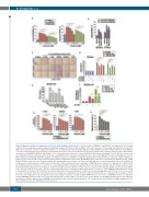
Figure 1. Maraviroc inhibits crosstalk between cHL cells and both MSCs and monocytes. (A) Percentages of BM-MSCs and AT-MSCs that migrated (in 5 h) through a fibronectin-coated Boyden chamber towards conditioned medium (CM) from L-1236 or KM-H2 cells, in the presence of increasing concentrations of maraviroc (MSCs were treated for 1 h prior to migration). (B) Effect of a neutralizing anti-CCL5 antibody (5 mg/mL) in cHL-conditioned medium on MSC migration. Transmigrated cells were revealed using a computer-interfaced GeniusPlus microplate reader (Tecan). Results are the mean and SD of three replicate wells from three independent experiments. (C) BM-MSCs, AT-MSCs and HL-MSCs (100 cells/well; 24-well plates) were cultured in RPMI-1640 medium containing 10% cHL CM. After 9 days, cells were fixed with methanol and stained with crystal violet. (D) BM-MSCs (500 cells/well; 96-well plates) were cultured in RPMI-1640 medium containing 20% cHL CM, with or without a neutralizing anti-FGF2 (1 mg/ml), anti-TGFb1 (2 mg/mL) or anti-TNFα (0.5 mg/mL) antibody. After 9 days, growth was evaluated using the MTT assay. Results are the mean and SD of three replicate wells from three independent experiments. (E) BM-MSCs were cultured for 6 days with 20% CM from L-1236, L-428, KM-H2, HDLM-2, and L-540 cells. Then, the medium was changed with fresh medium, and 3 days later MSC CM was recovered and quantified for CCL5 by ELISA. All samples were tested in triplicate; conditioned media from three different experiments were evaluated. (F) BM-MSCs were cultured for 6 days with 20% CM (from KM- H2 or HDLM-2 cells) in the presence or absence of a neutralizing anti-TNFα antibody (0.5 mg/mL). Then, the medium was changed and after 3 days CCL5 was quan- tified by ELISA. All samples were run in triplicate; conditioned media from three different experiments were evaluated. (G) Clonogenic growth. L-1236 (103/mL), HDLM- 2 (5 × 10²/mL), L-540 (2.5 × 10²/mL) cells were cultured in methylcellulose-containing medium in the absence or presence of 5% E-BM-MSC CM and with increasing concentrations of maraviroc. After 14 days of incubation, plates were observed under phase contrast microscopy and aggregates with 40 cells or more were scored as colonies (8 replicate wells). Each experiment was done in triplicate; conditioned media from three different experiments were evaluated. (H) Percentage of CD14+ monocytes that migrated (in 1 h) through fibronectin-coated Boyden chambers towards medium (RPMI-1640 plus 10% FCS) or E-BM-MSC CM. Prior to migration towards E-BM-MSC CM, monocytes were pretreated with maraviroc (0.1-100 mM) for 1 h. Results are means and SD of transmigrated monocytes for three different experiments. AT-MSCs: Adipose Tissue; BM-MSCs: Bone Marrow; cHL: classical Hodgkin lymphoma; CM: conditioned medium; HL-MSCs: Hodgkin lymphoma; MSC: Mesenchymal stromal cells; E-BM-MSCs: tumor Educated.
566
haematologica | 2019; 104(3)
