
Page 161 - 2019_03-Haematologica-web
P. 161
Maraviroc inhibits cHL crosstalk and xenograft growth
Results
Maraviroc inhibits crosstalk between cHL cells and both MSCs and monocytes
Conditioned medium from L-1236 and KM-H2 cHL-derived cell lines stimulated the migration of BM-MSCs (Figure 1A, left) and AT-MSCs (Figure 1A, right). Since MSCs express CCR5 (Online Supplementary Figure S1A)35 and cHL cells secret CCL5,7 we inves- tigated whether this chemokine is directly involved in MSC migration. Addition of the CCR5 antagonist maraviroc (Figure 1A) or a neutralizing anti-CCL5 antibody (Figure 1B) significantly reduced the MSC migration induced by cHL-conditioned medi um. Growth of MSCs from different sources (bone marrow, adi-
pose tissue, and cHL lymph nodes) increased in the presence of cHL-conditioned medium in a dose-dependent manner (Figure 1C and Online Supplementary Figure S1B). This effect was partially mediated by FGF2, TGFb1 and TNFα secreted by cHL cells, since addition of antibodies against these growth factors significantly, but incompletely, reduced growth (Figure 1D). cHL-conditioned medium almost totally abolished MSC senescence induced by serum starvation (Online Supplementary Figure S1C) and reduced apoptosis induced by doxorubicin treatment (Online Supplementary Figure S1D). To survive and proliferate, cancer cells not only recruit but also shape or “educate” normal cells.11 BM-MSCs released very low amounts of CCL5 under normal culture conditions (Figure 1E). After being cultured with cHL-conditioned medium, thereby
A
B
C
D
E
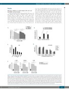
Figure 2. cHL cells induce monocyte migration and proliferation. (A) Percentages of monocytes that migrated (in 1 h) through fibronectin-coated Boyden chambers towards conditioned medium (CM) from L-1236 and L-428 cells, in the presence of increasing concentrations of maraviroc (monocytes were treated for 1 h prior to migration). (B) Effect of a neutralizing anti-CCL5 antibody (5 mg/mL) in cHL-conditioned medium on monocyte migration. Results are means and SD of three replicate wells from three independent experiments. (C) cHL cells (2x10⁵ cells/mL) were cultured for 3 days before CM was collected and tested for M-CSF by ELISA. All samples were run in triplicate; conditioned media from three different experiments were evaluated. (D) Monocytes (2.0x10⁴ cells/well; 96-well flat-bottomed plates) were exposed to increasing concentrations (percentage, v/v) of cHL CM. After 9 days, monocyte growth was evaluated using the MTT assay. Results are mean and SD of three experiments. (E) Clonogenic growth in methylcellulose-containing medium. L-1236 (103/mL), HDLM-2 (5x10²/mL), L-540 (2.5x10²/mL) cells were cultured in the absence or presence of 5% (v/v) E-monocyte CM, with increasing concentrations of maraviroc. After 14 days of incubation, plates were observed under phase contrast microscopy and aggregates with 40 cells or more were scored as colonies (8 replicate wells). Each experiment was done in triplicate; conditioned media from three different experiments were evaluated. cHL: classical Hodgkin lymphoma; CM: conditioned medium, (E)-monocytes: tumor Educated.
haematologica | 2019; 104(3)
567
