
Page 163 - 2019_03-Haematologica-web
P. 163
Maraviroc inhibits cHL crosstalk and xenograft growth
A
BC
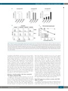
Figure 3. Monocyte conversion towards immunosuppressive E-monocytes by cHL cells. (A) Purified CD14+ monocytes were cultured for 6 days in the absence or presence of 20% conditioned medium (CM) from L-1236, L-428, and L-540 cells (to convert monocytes into E-monocytes), then washed and cultured for another 72 h in fresh medium. This E-monocyte CM was recovered for IL-10, CCL17, and TGFb ELISAs. Results are means and SD of three independent experiments. (B) Representative flow cytometric histograms showing surface expression of CD206 and PD-L1 and intracellular expression of IDO in monocytes (control) and E-mono- cytes (treated with cHL CM). (C) E-monocyte CM was tested for immunosuppressive activity. Phytohemagglutinin (PHA)-activated lymphocytes were treated with increasing concentrations (percentage, v/v) of monocyte CM and E-monocyte CM. After 72 h, growth was assayed using the Cell Proliferation ELISA, BrdU. All samples were tested in triplicate; conditioned media from three different experiments were evaluated. cHL: classical Hodgkin lymphoma; CM: conditioned medium, (E)-mono- cytes: tumor Educated.
assembling of cHL, HL-MSCs and monocytes into heterospher- oids (Figure 4E). It also reduced the total number of viable cells in the heterospheroids (Figure 4F). Considering this reduced viability, and the finding that maraviroc synergized with doxorubicin (Table 1), we applied drug combinations to the heterospheroids and found that doxorubicin and maraviroc exerted synergistic activity (combination index <1) against heterospheroids too (Online Supplementary Figure S6B). When cHL cells (CD30+) were recovered from heterospheroids by trypsinization and purification with anti-CD30 beads, fewer viable tumor cells were recovered from maraviroc-treated than from untreated heterospheroids (Online Supplementary Figure S6C). The cells from treated heteros- pheroids produced fewer colonies (Online Supplementary Figure S6D) and were proportionately more in G1 than in G2M phase than untreated cells (Online Supplementary Figure S6E).
Maraviroc slows the growth of cHL tumor xenografts and reduces infiltrated TAMs
To analyze the anticancer activity of maraviroc in vivo, we stud- ied L-540 tumor cell xenografts in female athymic nude mice, treated every day with an intraperitoneal injection of 10 mg/kg maraviroc or vehicle. By day 12, untreated tumors had grown to a mean volume of 880 mm3 (SD = 88 mm3), whereas maraviroc- treated tumors were more than 50% smaller (435±75 mm3; P<0.0001, Student’s t-test; Figure 5A and Online Supplementary
Figure S7A). Maraviroc treatment was not toxic as the animals were normal on physical inspection and had similar weight to untreated animals (Figure 5B). Maraviroc-treated mice had signifi- cantly better “survival” (i.e., tumor volume <800 mm3) than untreated mice (P=0.002, log rank test) (Online Supplementary Figure S7B). Since maraviroc inhibited the migration of monocytes in vitro, we evaluated whether similar activity was also detectable in vivo by examining infiltrating TAMs (CD68+) in L-540 tumor xenografts. Immunofluorescence analysis of CD30 on tissue sec- tions showed no difference between untreated and maraviroc- treated mice (Online Supplementary Figure S7C). However, the ani- mals differed substantially in staining for CD68, a marker of TAM infiltration, which was almost completely absent in maraviroc- treated mice (Figure 5C-D and Online Supplementary Figure S7D).
Similar results were obtained when male NSG mice were inject- ed with L-428 tumor cells (Figure 5E-H). In particular, maraviroc treatment reduced xenograft growth by about 60% (Figure 5E), without weight loss (Figure 5F), and it reduced CD68 staining by 75% (Figure 5G-H and Online Supplementary Figure S7D).
High CCL5 expression positively correlates with CD68 and poor survival
To confirm our in vitro and in vivo results, we studied cHL tissues from 65 patients (Online Supplementary Table S1). cHL tissues had a median CCL5 expression level of 12% positive pixels (range, 0%-
haematologica | 2019; 104(3)
569
