
Page 166 - 2019_03-Haematologica-web
P. 166
N. Casagrande et al.
We found that conditioned medium from cHL cells increased the growth of MSCs from different sources, and maraviroc decreased MSC recruitment by cHL cells. Thus, the development of fibrosis in cHL tissues could be explained by recruitment of MSCs by cytokines, including CCL5, secreted by primary tumors and expansion or acti- vation by FGF2, TGFb or TNFα secreted by tumor cells. Moreover, our findings that cHL education of MSCs (E-MSCs) increased the secretion of CCL5 and that mar- aviroc reduced monocyte recruitment by E-MSC-condi- tioned medium suggest an active role of E-MSCs in TME formation. In this perspective, MSCs of the cHL TME not only may down-regulate anti-tumor immune responses
through NKG2D-NKG2DL interactions,36,37 but may also enhance the number of infiltrated TAMs by secreting CCL5 and, consequently, supporting tumor progression.
Macrophages seem to be involved in the pathogenesis of cHL, since high levels of TAMs as well as the absolute monocyte count in the blood correlate with an unfavor- able clinical outcome.17,20,21,38 Despite the importance of monocyte levels in TME, their education (i.e., condition- ing by tumor cells) seems an essential prerequisite for their pro-tumor activity.39,40 Recently, it was demonstrated that conditioned medium from cHL cell lines induced an immunosuppressive phenotype in macrophages obtained by pre-cultivation of monocytes with M-CSF or GM-
A
BC
P=0.0072
HR (low-med/hi)=0.015
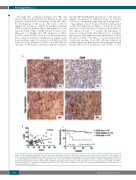
Figure 6. CCL5 expression positively correlates with CD68 expression in human cHL tissues and with lower progression-free survival in cHL patients. (A) Representative photomicrographs of cHL tissues stained for CCL5 and CD68 (marker of macrophages/monocytes). Top row, a case with low expression; bottom row, a case with high expression. (B) Correlation of expression levels of CCL5 and CD68 in 65 cHL patients. Spearman r=0.251; P=0.0487. (C) Kaplan-Meier survival plot for 5-year progression-free survival in cHL patients, subdivided according to the percentage of CCL5 positivity (low, 0%-10%; medium, 11%-20%; high, 21%-100%). Patients with high CCL5 expression had worse survival (P=0.0072, HR=0.015 vs. low-medium levels). cHL: classical Hodgkin lymphoma; HR: hazard ratio; r: Spearman correlation coefficient.
572
haematologica | 2019; 104(3)
