
Page 134 - 2019_03-Haematologica-web
P. 134
F. Portale et al.
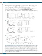
approximately 25% in CXCL12-driven migration (Figure 6B). This effect on CD34+ cell migration was not due to an ActivinA-mediated regulation of the CXCL12 chemokine receptors, CXCR4 and CXCR7, as demonstrated by flow cytometry analysis of both membrane-bound receptors and intracellular pool (Figure 6C). In addition, ActivinA pretreatment significantly decresed free cytosolic Ca2+ of CD34+ cells after the addition of CXCL12 in 2 out of 3
independent experiments (Figure 6D). Overall, these data suggest that leukemic cells could displace healthy hematopoietic stem cells from their niches through an ActivinA-mediated mechanism.
Acute lymphoblastic leukemia-mesenchymal stromal cells secrete high amounts of ActivinA
Finally, we focused our attention on the capacity of the
A
BC
D
Figure 6. ActivinA impaired CXCL12-driven migration of healthy CD34+ cells. (A) Expression of ActivinA receptors ALK2, ALK4, ACVR2A and ACVR2B was quantified in both cord blood (CB)-CD34+ and bone marrow (BM)-CD34+ cells by qRT-PCR. Data are presented as mRNA fold change of ActivinA receptors normalized to GAPDH mRNA (endogenous control). DAUDI cell line was employed as calibrator because of its low expression of ActivinA receptors (www.proteinatlas.org). (B) CB-CD34+ cells (top) and BM-CD34+ (bottom) were pretreated or not with ActivinA (100 ng/mL) for 24 hours (h) and allowed to migrate through 5 mM pores in Transwell cham- bers toward CXCL12 (100 ng/mL) for 1 h. The graphs represent one representative experiment (CB-CD34+ n=5, BM-CD34+ n=3). Each box plot shows the median and the mean (+) of the percentage of migrated cells, and extends from the lowest to the highest value. *P<0.05: Mann-Whitney test. (C) The extracellular and intra- cellular levels of CXCR4 and CXCR7 were analyzed in cells either treated (black line) or not (gray line) with ActivinA for 24 h by flow cytometry. Data from one repre- sentative experiment out of three are shown. (D) CB-CD34+ cells were cultured for 24 h in the presence or absence of ActivinA (100 ng/mL). Cells were loaded with Fluo-4 NW and free cytosolic Ca2+ changes were measured by FACS. Background was recorded for 30 seconds (s) and signal upon CXCL12 addition was registered for an additional 90 s. The black line represents results obtained from ActivinA-treated cells, while the gray line represents untreated cells. The results are represen- tative of three independent experiments. *P<0.05: Mann-Whitney test (Ppeak R2: comparison between MFI peak range 2 in treated vs. untreated cells; Pmean R2: comparison between MFI mean range 2 in treated vs. untreated cells).
540
haematologica | 2019; 104(3)
