
Page 132 - 2019_03-Haematologica-web
P. 132
F. Portale et al.
A
B
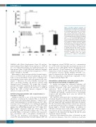
Figure 4. ActivinA enhanced leukemic cell responsiveness to CXCL12. (A) CXCL12 bone marrow plasma levels were assessed by ELISA in 46 healthy donors (HDs) and 70 B-cell pre- cursor-acute lymphoblastic leukemia (BCP- ALL) patients at the onset of the disease. Each box plot shows the median and the mean (+) and extends from the lowest to the highest value. ****P<0.0001: Mann-Whitney test. (B) 697 cells were pretreated with ActivinA (50 ng/mL) for 24 hours (h) and then incubated for 4 h in Transwell chambers toward decreasing concentration of CXCL12 (100-10-1 ng/mL). Each box plot shows the median and the mean (+) of the percentage of migrated cells and extends from the lowest to the highest value. The graphs represent the results of six inde- pendent experiments. *P<0.01: ActivinA-treat- ed versus untreated 697 cells, Wilcoxon matched-pairs signed rank test; #P<0.05; ##P<0.01: comparison with 100 ng/mL CXCL12-induced migration, Mann-Whitney test.
NALM-6 cells (Online Supplementary Figure S6) and pri- mary leukemic blasts (Figure 3D) in response to CXCL12 in a dose-dependent manner. In 3 different patients, we demonstrated that 10 mM SB431542 inhibited ActivinA stimulatory effect on CXCL12-driven migration of 78.8% (range: 74.5-84.0%; P<0.05).
Interestingly, it has been reported that ActivinA expres- sion is associated with an invasive phenotype in several types of cancer, including ovarian cancer, esophageal ade- nocarcinoma, breast cancer, and oral squamous cell carci- nomas.12-15 Therefore, we tested whether ActivinA was able to modulate leukemic cell invasive capacity using Matrigel-coated Transwells. We found that ActivinA increased the ability of primary BCP-ALL cells to migrate through a complex matrix in the presence of CXCL12 (Figure 3E), with a 2-fold increase compared to the untreated condition (P<0.05).
ActivinA enhanced leukemic cells responsiveness to low levels of CXCL12
CXCL12 reduction is one of the microenvironmental alterations occurring in the leukemic BM, as observed in both mice models and leukemic patients,18,19 which is asso- ciated with impairment of normal hematopoiesis.5 Here, in a large cohort of 70 patients, we confirmed a significant reduction of approximately 6 times the CXCL12 of BM plasma level in BCP-ALL patients (mc: 77.7, range: 15.7- 488.9 pg/mL) compared to HDs (mc: 476.8, range: 99.1- 1763 pg/mL, n=46) (P<0.0001) (Figure 4A). To test the potential ability of ActivinA to increase the responsive- ness of leukemic cells to suboptimal concentrations of CXCL12, we performed dose-response chemotaxis assays. We demonstrated that ActivinA enhanced 697 cell
line migration toward CXCL12 used at a concentration 10- or 100-fold lower than that classically used in in vitro migration assays (100 ng/mL). Indeed, ActivinA pretreat- ment induced a 10-fold increase in the CXCL12-driven chemotaxis toward 10 ng/mL CXCL12 (P<0.05) and a 7- fold increase toward 1 ng/mL CXCL12 (P<0.05), com- pared to untreated cells, that showed a responsiveness to these low chemokine concentrations comparable to that of empty medium (Figure 4B).
Intracellular calcium levels and actin polymerization were increased by ActivinA in leukemic cells
To further investigate the enhanced leukemic cell responsiveness to CXCL12, we first evaluated whether ActivinA treatment could affect chemokine receptor expression. Flow cytometry analysis of CXCL12 chemokine receptors on 697 cells showed that the levels of CXCR4 and CXCR7, evaluated both as extracellular receptors and intracellular pool, were not affected by ActivinA (Figure 5A).
We, therefore, performed flow cytometry analysis to determine the effect of ActivinA on the intracellular calci- um level of BCP-ALL cells. Our data indicated that both in 697 cells (Figure 5B) and in primary BCP-ALL cells (Figure 5C) the basal intracellular calcium content was increased in ActivinA-pretreated cells as compared to untreated cells (mean range 1; P<0.05). Interestingly, on addition of CXCL12, ActivinA-treated cells showed a further signifi- cant increase in the concentration of free cytosolic Ca2+ compared to the untreated cells (Figure 5B and 5C, peak and mean range 2; P<0.05).
Moreover, we evaluated the effect of ActivinA on actin cytoskeleton dynamics. Since the conversion of globular
538
haematologica | 2019; 104(3)
