
Page 131 - 2019_03-Haematologica-web
P. 131
ActivinA enhances BCP-ALL cell migration
A
B
CDE
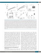
Figure 3. ActivinA enhanced cell motility, migration and invasion of leukemic cells. (A) Leukemic cells were treated or not with ActivinA and then tracked for 24 hours (h) by time-lapse microscopy. Dead cells were excluded using PI staining. Data represent the mean±Standard Error of Mean (SEM) of the migrated distance over time of three independent experiments. The distance migrated after 24 h by all tracked cells was compared between ActivinA-stimulated (gray) and not stimulated (white) cells. (Left) 697 cells treated with 50 ng/mL ActivinA. (Right) B-cell precursor-acute lymphoblastic leukemia (BCP-ALL) primary blasts treated with 100 ng/mL ActivinA. (B) Chemotaxis assay was performed using 697 cells stimulated with ActivinA for 24 h (50 ng/mL) and allowed to migrate toward CXCL12-containing medium (100 ng/mL) for 4 h (5 mM pore Transwell). Each box plot shows the median and the mean (+) of the percentage of migrated cells and extends from the lowest to the highest value. The graphs represent the results of six independent experiments. The percentage of migrated cells was determined as described in the Online Supplementary Methods. (C) Primary BCP-ALL cells from 13 patients were exposed to ActivinA (100 ng/mL) for 24 h and employed for chemotaxis assays toward CXCL12-containing medium (100 ng/mL). The average percentage of cells migrated after 1 h of culture is shown. (D) Chemotaxis assay was performed using BCP- ALL primary cells pretreated for 1 h with SB431542 or vehicle (DMSO) before the stimulation with or without ActivinA for 24 h (100 ng/mL). Cells were allowed to migrate toward CXCL12-containing medium (100 ng/mL) for 1 h (5 mM pores Transwell). The percentage of migrated cells was determined as described in the Online Supplementary Methods. The average percentage of inhibition±SEM is represented (one out of three representative experiments). (E) Primary BCP-ALL cells pretreat- ed or not with ActivinA for 24 h (100 ng/mL) were allowed to migrate through Transwell inserts (8 mM pores) coated with a Matrigel-barrier (1 mg/mL) for 24 h in the presence of CXCL12 (100 ng/mL) in the lower chamber. Each box plot shows the median and the mean (+) of the percentage of invaded cells and extends from the lowest to the highest value. The graphs represent the results of 3 different patients. *P<0.05; ***P<0.001; ****P<0.0001; Wilcoxon matched-pairs signed rank test (A-C); *P<0.05: comparison with vehicle (0 mM), Mann-Whitney test; #P<0.05: comparison with 10 mM SB431542, Mann-Whitney test (D); *P<0.05: Mann- Whitney test (E).
the expression of several genes linked to Ca2+ homeostasis (ATP2B2, ATP2B4), Ras pathway activation (VAV3), and cell motility and movement regulation (ARHGAP25, CORO1A, DOCK4, LCK, PTPRC). Data obtained in qRT- PCR on 697 cells were highly concordant with microarray data (Online Supplementary Figure S3B). Raw data are shown in Online Supplementary Figure S4. In addition, qRT-PCR analyses of the above-mentioned genes were performed on 7 primary BCP-ALL patients' samples stim- ulated or not with ActivinA (Online Supplementary Figure S5). Among them, 3 presented the t(1;19) (gray dots) typ- ical of the 697 cell line. Interestingly, four of the tested genes (ARHGAP25, CORO1A, DOCK4 and PTPRC) were significantly modulated in at least one stimulation time point (Online Supplementary Figure S5B) by ActivinA in more than 70% of patients, similarly to our observations in 697 cells. It is worth noting that the t(1;19) patients showed a modulation of the ATP2B4, VAV3 and LCK genes (Online Supplementary Figure S5B) more similar to 697 cells than the translocation negative ones.
ActivinA increased random motility, chemotaxis and invasion of B-cell precursor-acute lymphoblastic leukemia cells
To test whether ActivinA was able to modulate BCP- ALL movement, we performed time-lapse microscopy
(TLM) analyses and migration assays. TLM studies showed that ActivinA was able to increase random motil- ity both in the 697 cell line (P<0.0001) (Figure 3A, left) and primary leukemic cells (P<0.0001) (Figure 3A, right).
It has been demonstrated that chemokines, and in par- ticular the CXCR4/CXCL12 axis, play a key role in the homing and retention of ALL cells within the BM niche. Therefore, we tested whether ActivinA was able to mod- ulate CXCL12-induced migration of leukemic cells using a Transwell-based migration assay. Notably, we found that ActivinA-pretreated 697 cells showed a significant increase in CXCL12-driven migration (P<0.05) (Figure 3B). Importantly, this finding was confirmed in primary leukemic cells obtained from the BM of 13 BCP-ALL patients collected at diagnosis (Figure 3C). The average percentage of primary unstimulated BCP-ALL blasts migrated in response to CXCL12 (mean: 15.6%, range: 1.9-43.5%) was significantly increased following ActivinA stimulation (mean: 22.9%, range: 4.1-61.1%) (P<0.001).
To ensure the specificity of the observed effect on CXCL12 mediated migration, we blocked ActivinA/Activin receptor axis by using SB431542, a well- characterized specific inhibitor of transforming growth factor-b superfamily type I Activin receptor-like kinase (ALK) receptors ALK4, ALK5, and ALK7.16,17 SB431542 inhibited the migration of ActivinA-pretreated 697,
haematologica | 2019; 104(3)
537
