
Page 133 - 2019_03-Haematologica-web
P. 133
ActivinA enhances BCP-ALL cell migration
A
B
C
D
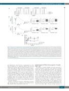
Figure 5. ActivinA increased CXCL12-induced calcium mobilization and actin polymerization. (A) The extracellular and intracellular levels of CXCR4 and CXCR7 were analyzed by flow cytometry in 697 cells treated (black line) or not (gray line) with ActivinA for 24 hours (h). Representative data are shown from three independent experiments. 697 cells (B) and B-cell precursor-acute lymphoblastic leukemia (BCP-ALL) primary blasts (C) were cultured for 24 h either in the presence or in the absence of ActivinA (50 ng/mL or 100 ng/mL, respectively). Cells were loaded with Fluo-4 NW and free cytosolic Ca2+ changes were measured by FACS. Background was recorded for 30 seconds (s) and signal upon CXCL12 addition was registered for an additional 90 s. The black line represents data obtained from ActivinA-treated cells; the gray line represents untreated cells. The arrow indicates CXCL12 addition. The box plots represent the mean fluorescence intensity (MFI) before (mean range 1) and after (mean range 2) the addition of CXCL12 and the maximum peak reached upon CXCL12 addition (peak range 2). Each box plot shows the median and the mean (+), and extends from the lowest to the highest value. The results are representative of one out of six independent experiments. (D) 697 cells were starved in low serum medium for 24 h and then stimulated or not with ActivinA (50 ng/mL) for an additional 24 h. Cells were then stained with AF647-phalloidin and MFI quantified by flow cytometry. Percentage of MFI change was defined as follows: (MFI after CXCL12 addition/ MFI before CXCL12 addition) x 100. Mean values (±Standard Error of Mean) of one out three independent experiments are represented in the graph. *P<0.05: Wilcoxon matched-pairs signed rank test (B and C). *P<0.05: Mann-Whitney test (D).
into filamentous actin (F-actin) is a prerequisite for site- directed migration, we analyzed whether the increased chemotactic response upon ActivinA treatment was asso- ciated with enhanced chemokine-induced actin polymer- ization. Pretreatment of 697 cells with ActivinA for 24 h resulted in a more prominent conversion of globular into F-actin starting from 15 seconds (s) after addition of CXCL12 (P<0.05) (Figure 5C). Notably, ActivinA-pretreat- ed cells maintained a higher amount of F-actin compared to untreated cells, even 180 s after CXCL12 stimulation (P<0.001). These data strongly support our GEP results highlighting the role of ActivinA as a modulator of several genes involved in cytoskeleton remodeling and regulation of calcium dynamics.
Results on migration, invasion, chemokine receptors, and calcium flux were confirmed in the NALM-6 BCP- ALL cell line (Online Supplementary Figure S7).
ActivinA impaired CXCL12-driven migration of healthy CD34+ cells
We further evaluated whether ActivinA promoted a selective advantage to BCP-ALL cells compared to healthy CD34+ cells. CB- and BM-derived CD34+ cells expressed both type I and type II Activin receptors, thus suggesting that they could both respond to this molecule (Figure 6A).
The effect of ActivinA on CXC12-driven chemotaxis of CD34+ cells was evaluated by Transwell-based migration assays. Surprisingly, we observed the opposite effect to that observed with leukemic cells. ActivinA pretreatment resulted in an average reduction of approximately 55% of CXCL12-driven chemotaxis, compared to untreated CB-CD34+ cells (Figure 6B). Of note, the regulation of cell viability did not account for the reduced chemotaxis (data not shown). These data were confirmed in BM-CD34+ cells derived from three HDs, with an average reduction of
haematologica | 2019; 104(3)
539
