
Page 130 - 2019_03-Haematologica-web
P. 130
F. Portale et al.
A
B
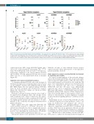
Figure 2. B-cell precursor-acute lymphoblastic leukemia (BCP-ALL) cells expressed type I and type II ActivinA receptors. (A) The expression of the type I ActivinA receptors ALK2 and ALK4 and the type II ActivinA receptors ACVR2A and ACVR2B was quantified in five leukemic cell lines (697, NALM-6, RS4;11, SUP-B15, REH) and (B) in bone marrow (BM) primary blasts from BCP-ALL patients by western blot (ALK2, n=12) and flow cytometry (ALK4, ACVR2A, ACVR2B, n=18). Data are pre- sented as the ratio of ALK2 to b-actin obtained by densitometric analysis and mean fluorescence intensity (MFI) for ALK4, ACVR2A and ACVR2B.
soluble factors (mc: 268.1, range: 62.5-842.8 pg/mL) and a cell-to-cell contact-mediated mechanism (mc: 777.9, range: 96.2-1456 pg/mL), with a 2.6 and a 7-fold increase, respectively, compared to the basal condition (P<0.001 and P<0.0001). Notably, primary BCP-ALL cells secreted either very low or even undetectable levels of ActivinA (Figure 1B).
Leukemic cells expressed ActivinA receptors
To determine whether BCP-ALL cells could be targets of ActivinA, the expression of Activin receptors was assessed on five leukemic cell lines (697, NALM-6, RS4;11, SUP- B15, REH) (Figure 2A) and eighteen primary BCP-ALL blasts by flow cytometry and western blot analyses (Figure 2B). Western blot images are shown in Online Supplementary Figure S1.
Both type I and type II Activin receptors were found to be expressed by all primary blasts and cell lines tested, with a markedly wide range of expression. The expres- sion level of ActivinA receptors in primary BCP-ALL cells was highly patient-specific and was shown to be inde- pendent of the commonly investigated leukemia-related genetic alterations.
Taken together, these data suggest that BCP-ALL cells could possibly respond to ActivinA. Moreover, we showed that ActivinA was able to significantly increase the expression of its type I receptors, thus suggesting a positive loop underlying the responsiveness of leukemic cells to ActivinA (Online Supplementary Figure S2).
Subsequent analyses were performed on the 697 and
NALM-6 cell lines as their different Activin receptor expression makes them representative of the high inter- patient variability observed.
Gene expression analysis revealed ActivinA involvement in regulating cell motility
For a more in depth analysis of the molecular changes induced by ActivinA in BCP-ALL cells, we performed gene expression profile (GEP) analysis of 697 cells upon stimu- lation with ActivinA for 6 h and 24 h. We found that 122 genes were differentially expressed in ActivinA-treated cells versus untreated cells after 6 h of stimulation (FDR<0.05) and that 151 genes were differentially expressed after 24 h of stimulation (FDR<0.05). Gene Ontology (GO) analysis of differentially expressed genes identified enriched GO categories (Online Supplementary Figure S3A) critically linked to cancerogenesis such as “reg- ulation of cell activation”, “positive regulation of antigen receptor-mediated signaling pathway”, “pathways in can- cer”, etc.
Interestingly, we also observed that ActivinA was able to influence migration-associated pathways, such as “cal- cium ion homeostasis and transport into cytosol", “PI3K/AKT activation”, “Ras signaling pathway”, “focal adhesion", suggesting its possible effect on leukemic cell motility. These data are in agreement with the recently recognized role of ActivinA in the regulation of cell migra- tion and invasion in the context of several solid malignan- cies.12-15 On the basis of this evidence, we first used qRT- PCR assays to validate the ActivinA-mediated changes in
536
haematologica | 2019; 104(3)
