
Page 136 - 2019_03-Haematologica-web
P. 136
F. Portale et al.
A
B
C
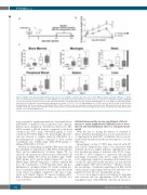
Figure 8. NALM-6 cells stimulated with ActivinA showed an increased ability to engraft in vivo in the bone marrow (BM), meninges and brain of NSG mice. (A) NSG mice received intravenous (i.v.) transfer of 1x10⁶ NALM-6 cells, previously cultured for 24 hours (h) in the presence or absence of ActivinA (50 ng/mL). (B) Weight loss was periodically monitored over two weeks after transplantation. The graph shows mean percentages±Standard Error of mean (SEM) of body weight change. Data from 12 mice/group in four independent experiments are shown. *P<0.05; **P<0.01: Mann-Whitney test. (C) Mice were sacrificed on days 11 and 14 after transplantation and the percentage of infiltrating hCD19+ hCD10+ leukemic cells was determined by flow cytometry in the BM, peripheral blood, spleen, liver, meninges and brain. Each box plot shows the median and the mean (+), and extends from the lowest to the highest value (n=12 mice/group, four independent exper- iments). *P<0.05: Mann-Whitney test.
assays revealed a significant induction of ActivinA release in BM-MSCs compared to their respective basal condition. Indeed, upon stimulation, ActivinA production by HD- MSCs reached a 28-fold increase compared to the basal condition (mc: 5713, range: 1446-14221 pg/mL vs. basal condition) (P<0.0001) (Figure 7A). Interestingly, the mole- cule was released to a higher extent by ALL-MSCs in a pro-inflammatory condition compared to their normal counterparts (mc: 10085, range: 2904-19776 pg/mL vs. inflamed HD-MSCs; P<0.01).
Notably, by mimicking an inflamed BM niche through the simultaneous stimulation of HD-MSCs with leukemic blasts and pro-inflammatory cytokines (Figure 7B), we showed a strong increase in the secretion of ActivinA both in the direct (Figure 7B, bottom, mc: 27860, range: 13150- 92391 pg/mL, n=17) and the indirect (Figure 7B, top, mc: 25409, range: 9050-65714 pg/mL) co-culture condition. Of note, the combination of both leukemic blasts and pro- inflammatory cytokines (Figure 7B, fourth column) pro- duced a synergistic induction of ActivinA, since the extent of the release was higher compared to the sum of sepa- rately used stimuli21 (Figure 7B, expected additive effect: fifth column=second+third columns; top: P<0.001; bot- tom: P<0.0001).
ActivinA increased the in vivo engraftment of B-cell precursor acute lymphoblastic leukemia cells to bone marrow and extramedullary sites in a xenograft mouse model
With the aim of testing the efficacy of ActivinA to induce leukemia dissemination in vivo, we performed a set of experiments in which 697 or NALM-6 cells in vitro pre- treated with ActivinA for 24 hours were injected (i.v.) into NSG mice.
Interestingly, on day +7, NSG mice injected with 697 cells (Online Supplementary Figure S10) pretreated with ActivinA showed a higher leukemic engraftment in the liver (median percentage of leukemic cells: 48.4%, range: 19.2-54.1%, n=9) compared to untreated cells (median percentage of leukemic cells: 27.0%, range: 10.9-43.2%, n=9), suggesting a migratory advantage in ActivinA-treat- ed cells. As expected from the literature, our data showed a high tropism of 697 cells for the liver.22,23 On the contrary, the percentages of leukemia engraftment in BM and in other leukemic target organs were modest and were com- parable between the two experimental groups.
To test a more physiological environment for leukemia and overcome the low engraftment of 697 in BM, we transplanted mice with NALM-6 cells that are known to
542
haematologica | 2019; 104(3)
