Page 188 - Haematologica-April 2018
P. 188
N. Wohmer et al.
of wt-VWF to SR-AI was also not altered upon incubation with ristocetin or botrocetin, indicating that binding does not require VWF to be in its platelet-binding conformation (data not shown). Interestingly, we detected little competi- tion between fragments (Figure 2D), suggesting that each domain binds to a distinct site within SR-AI, which at best overlap with each other.
SR-AI function is partially cation-dependent,25 and we therefore assessed binding of VWF in the presence of EDTA. This revealed that binding of VWF to SR-AI was reduced by 88±6% in the presence of EDTA (Figure 2E). Specificity was further investigated by testing the effect of monoclonal antibodies targeting VWF. We identified two antibodies (i.e. Mab723 and Mab540, directed against the A1 and D4 domain, respectively) that each partially inter-
fered with the binding of VWF to sSR-AI (48±2% and 47±2% inhibition by Mab723 and Mab540, respectively; n=3) (Figure 2E). A combination of both antibodies reduced binding by 72±4% (n=4; Figure 2E). Thus, puri- fied VWF interacts in a specific manner with SR-AI and several domains of the VWF molecule contribute to this interaction.
von Willebrand factor binds to cellular SR-AI
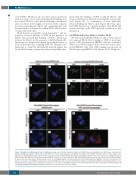
We next tested whether VWF was able to bind cell-sur- face exposed SR-AI. First, binding of VWF to non-trans- fected or SR-AI-transfected HEK293 cells was examined. Whereas no VWF staining could be detected on non-trans- fected HEK293 cells, clear VWF staining was present on SR-AI-expressing HEK293 cells (Figure 3A-D). High-reso-
ACE
BD
F
GIK
H
JL
732
Figure 3. Binding of van Willebrand factor to SR-AI-expressing cells. (A-D) Non-transfected (A&C) and hSR-AI-transfected HEK293-cells (B&D) were incubated with purified pd-VWF (10 mg/mL). hSR-AI and bound VWF were probed using polyclonal anti-hSR-AI (red, A&B) and anti-VWF antibodies (green, C&D). Images were obtained via widefield microscopy (objective 40x; scale bars: 10 mm). (E-F) Spinning disk microscopy images (objective 63x; scale bars: 5 mm, z-depth 0.5 mm) of hSR-AI-transfected HEK293-cells incubated with pd-VWF (10 mg/mL). Cells were probed for VWF and hSR-AI (E, green and red, respectively) and for VWF and EEA-1 (F, green and red, respectively). Arrows indicate areas of overlapping signals. (G-J) Non-transfected and SR-AI-transfected HEK293 cells were incubated in the absence or presence of pd-VWF (10 mg/mL). Association with SR-AI was detected using Duolink-PLA analysis by combining anti-VWF and anti-SR-AI antibodies. (K-L) THP1-derived macrophages were incubated in the absence or presence of pd-VWF (10 mg/mL). Association with SR-AI was detected using Duolink-PLA analysis by combining anti-VWF and anti-SR-AI antibodies. Panels G to L: objective 63x; scale bars: 10 mm.
haematologica | 2018; 103(4)


