
Page 186 - Haematologica-April 2018
P. 186
N. Wohmer et al.
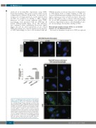
addressed in Duolink-PLA experiments using THP1- derived macrophages. This assay is positive when a recep- tor/ligand pair is within a 40-nm radius. As expected, no red spots above background levels could be detected when wt-VWF was analyzed for binding to LRP1, whereas numerous red spots became apparent when THP1- macrophages were incubated with the VWF-type 2B mutant VWF/p.V1316M (Figure 1A-C). This difference was confirmed when quantifying fluorescent signals (Figure 1D; n=5). In control experiments, we also stained THP1-macrophages in a classical manner for the presence of VWF. Interestingly, for those cells incubated with pd-
VWF, the majority of cells proved positive for the presence of VWF (Figure 1E,F). A similar binding of VWF was observed when human macrophages derived from circulat- ing blood precursors were used (data not shown). These data indicate that VWF is able to interact with macrophages also in an LRP1-independent manner, even under static conditions. Apparently, receptors other than LRP1 are pres- ent on macrophages that mediate binding of VWF.
Macrophage-specific receptor SR-AI as a potential receptor for von Willebrand factor
In search for alternative receptors for VWF, we explored
D
E
ABC
F
Figure 1. von Willebrand factor can bind to macrophages independently of LRP1. (A-D) THP-derived macrophages were incubated in the absence or presence of culture-medi- um containing wt-VWF or VWF/p.V1316M. Association with LRP1 was detected using Duolink-PLA analysis by combining anti-VWF and anti-LRP1 antibodies. Representative images are shown in panels A-C (objective 63x). Quantification of flu- orescent signals is shown in panel D. Data represent mean±SD (n=5 microscopic fields; 2-7 cells/field). Statistical analysis involved one-way analysis of variance followed by the Tukey multiple comparison test. (E, F) Classical immunefluo- rescence staining of THP1-derived macrophages incubated in the (E) absence or (F) presence of purified pd-VWF. Bound VWF was probed using monoclonal anti-VWF antibodies (objective 40x). Scale bars represent 10 mm in all panels.
730
haematologica | 2018; 103(4)
