
Page 187 - Haematologica-April 2018
P. 187
SR-AI contributes to VWF clearance
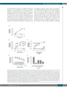
the option that SR-AI could be one of these receptors. SR- AI (also known as SCARA1 or CD204) is specifically expressed on macrophages and is structurally related to SCARA5, an epithelial cell-specific receptor that was iden- tified in genome-wide association studies to be linked to VWF plasma levels.24 We first analyzed binding of VWF to the soluble extracellular domain of SR-AI (sSR-AI) in an immunosorbent-based assay. Whereas no binding of VWF to albumin-coated control wells was observed, VWF dis- played saturable and dose-dependent binding to immobi- lized sSR-AI (half-maximal binding 3.5±1.2 mg/mL corre- sponding to 14±5 nM; n=5) (Figure 2A). We next assessed
A
BC
the capacity of sSR-AI to interact with various immobi- lized VWF fragments. sSR-AI bound dose-dependently to each of the three VWF fragments tested (the recombinant D’D3 and A1-A2-A3 regions and the D4/Fc fragment) (Figure 2B), suggesting that the interaction with SR-AI involves different regions of the VWF molecule. Further experiments showed that the A1 domain mediated bind- ing of the A1-A2-A3 fragment to sSR-AI, while both the A2 and A3 domains were incapable of associating with sSR-AI (Figure 2C). In addition, incorporation of the VWD-type 2B mutation VWF/p.V1316M left binding of the A1-domain to sSR-AI unaffected (Figure 2C). Binding
DE
Figure 2. von Willebrand factor interacts with SR-AI via multiple interactive sites. (A) Wells coated with recombinant human sSR-AI (closed circles) or bovine serum albumin (BSA) (open circles) were incubated with various concentrations of purified pd-VWF (0-5 mg/mL). Bound VWF was probed with peroxidase-labeled polyclonal anti-VWF antibodies. (B) Wells coated with recombinant VWF-fragments (closed squares: A1-A2-A3 domain; closed circles: D4-domain fused to Fc fragment; open squares: D’-D3 domains) or BSA (control; open circles) were incubated with various concentrations of sSR-AI (0-5 mg/mL). Bound sSR-AI was probed using biotinylated polyclonal anti-SR-AI antibodies followed by peroxidase-labeled streptavidin. (C) Wells coated with sSR-AI were incubated with various concentrations (0-5 mg/mL) of recombinant VWF-fragments (closed circles: A1-Fc; gray squares: A1-Fc/p.V1316M; gray circles: A2:Fc; open squares: A3-Fc). Bound fragments were probed using peroxidase-labeled polyclonal anti-human Fc antibodies. Control represents binding of A1-Fc to BSA-coated wells (control; open circles). Other fragments gave similar background signals. (D) sSR-AI-coated wells were incubated with recombinant D4-Fc fragment in the presence of various concentrations of recombinant A1-A2-A3 fragment (open circles) or recombinant D’D3 fragment (gray circles). Alternatively, sSR-AI-coated wells were incubated with recombinant A1-Fc fragment in the pres- ence of various concentrations of recombinant D’D3 fragment (closed squares). Bound fragments were probed using peroxidase-labeled polyclonal anti-human Fc antibodies. (E) sSR-AI-coated wells were incubated with VWF (2.5 mg/mL) in the absence or presence of EDTA (10 mM), monoclonal anti-VWF antibody MAb723 or MAb540 (25 mg/mL) or with both antibodies simultaneously (25 mg/mL each). Bound VWF was probed using peroxidase-labeled polyclonal anti-VWF antibodies. For all panels, bound antibodies were detected via TMB-hydrolysis. Data represent mean±SD (n=3-5).
haematologica | 2018; 103(4)
731
