
Page 190 - Haematologica-April 2018
P. 190
N. Wohmer et al.
von Willebrand factor propeptide to antigen ratio is lower in SR-AI-deficient mice than in wild-type and macLRP1-deficient mice
To assess the physiological relevance of SR-AI in regu- lating VWF clearance, we opted to express human VWF in wild-type, macLRP1- and SR-AI-deficient mice via hydro- dynamic gene transfer, and to determine the ratio between VWFpp and VWF:Ag, a measure of VWF clear- ance. As reported previously,9 VWFpp/VWF:Ag ratios were slightly, but significantly reduced in macLRP1-defi- cient mice compared to wt-mice (1.3±0.1 versus 1.1±0.1 for wt- and macLRP1-deficient mice, respectively; n=8-9; P=0.0114) (Figure 5). This confirms that LRP1 contributes to a modest extent to the clearance of VWF. Interestingly, VWFpp/VWF:Ag levels were even further reduced in SR- AI-deficient mice: 0.6±0.2 versus 1.3±0.3 (n=9-14; P<0.0001) (Figure 5). VWF is apparently cleared less rapid- ly in SR-AI-deficient mice than in macLRP1-deficient mice. This suggests that SR-AI plays a more dominant role than that of LRP1 in basal VWF clearance.
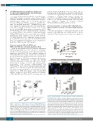
Clearance mutants VWF/p.R1205H and VWF/p.S2179F show enhanced binding to SR-AI
Given the involvement of the D’D3 and D4 domains in SR-AI binding (Figure 2), it was of interest to investigate whether clearance mutations in these domains affect the interaction with SR-AI. We first analyzed binding of VWF/p.R1205H (the Vicenza variant with a mutation in the D3 domain) and VWF/p.S2179F (with a mutation in the D4 domain) to sSR-AI in an immunosorbent assay. Whereas the interactions of type 2B mutant VWF/p.V1316M and wt-VWF with sSR-AI were similar (half-maximal binding at 3.1±0.7 and 3.6±0.9 mg/mL, respectively), both mutants VWF/p.R1205H and VWF/p.S2179F proved more efficient in interacting with SR-AI (1.7±0.3 and 2.3±0.4 mg/mL; P=0.0124) (Figure 6A). We then visualized binding of both mutants to SR-AI expressed on THP1-macrophages using Duolink-PLA analysis. Bright red spots were observed for mutants VWF/p.R1205H and VWF/p.S2179F, indicating that both
mutants interact with SR-AI at the macrophage cell sur- face (Figure 6B-D). Quantitative analysis revealed that flu- orescence was significantly increased for both mutants compared to wt-VWF. VWF surface coverage was 3.2±0.9% for wt-VWF, 8.7±2.4% for VWF/p.R1205H and 11.6±2.1% for VWF/p.S2179F (Figure 6E). These data indi- cate enhanced binding of clearance mutants VWF/p.R1205H and VWF/p.S2179F to SR-AI.
Increased clearance of mutants VWF/p.R1205H and VWF/p.S2179F is partially corrected in SR-AI-deficient mice
We next investigated to what extent clearance of the mutants VWF/p.R1205H and VWF/p.S2179F is SR-AI- dependent. Hydrodynamic gene transfer was applied to
A
BCD
E
Figure 6. Enhanced binding of von Willebrand factor mutants p.R1205H and p.S2179F to SR-AI. (A) Wells coated with sSR-AI were incubated with various concentrations of non-purified recombinant VWF (0-5 mg/mL). Closed circles: wt- VWF; open squares: p.V1316M; open diamonds: p.S2179F; closed diamonds: p.R1205H. Open circles represent binding of wt-VWF to bovine serum albumin- coated wells. Mutants gave similar background signals. Bound VWF was probed with peroxidase-labeled polyclonal anti-VWF antibodies. All mutants reacted sim- ilarly with these polyclonal antibodies. Data represent mean±SD (n=3). (B-E) THP1-derived macrophages were incubated in the absence or presence of non- purified recombinant wt-VWF (B) or mutants VWF/p.R1205H (C) or VWF/p.S2179F (D). Association with SR-AI was detected using Duolink-PLA analysis by combining anti-VWF and anti-SR-AI antibodies (Objective 63x; scale bars 10 mm). (E) Quantification of fluorescent signals. Data represent mean±SD (n=5 microscopic fields; 2-5 cells/field). Statistical analysis involved one-way analysis of variance followed by the Tukey multiple comparison test.
Figure 5. Deficiency of SR-AI results in decreased VWFpp/VWF:Ag ratios.
Human-wt-VWF was expressed in macLRP1+- and SR-AI-expressing mice and in macLRP1-deficient and SR-AI-deficient mice following hydrodynamic gene trans- fer. Four days after injection, plasma samples were prepared for the analysis of VWFpp and VWF:Ag. Assays for VWFpp and VWF:Ag quantify only human VWF expressed via hydrodynamic gene transfer, and do not cross-react with endoge- nous murine VWF. VWFpp/VWF:Ag ratios for each individual mice included in the study are plotted. Data from macLRP1-mice and SR-AI-mice were compared in a pairwise manner using a two-tailed Student t-test.
734
haematologica | 2018; 103(4)
