
Page 191 - Haematologica-April 2018
P. 191
SR-AI contributes to VWF clearance
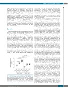
express human VWF, VWF/p.R1205H and VWF/p.S2179F in control mice and SR-AI-deficient mice, and the VWFpp/VWF:Ag ratio was determined. VWFpp/VWF:Ag ratios were markedly increased for both mutants in con- trol mice [2.9±0.2 (n=8) and 4.4±0.5 (n=7), for VWF/p.R1205H and VWF/p.S2179F, respectively; P<0.001 compared to wt-VWF] (Figure 7), confirming that both mutations induce increased clearance of VWF. When expressed in SR-AI-deficient mice, a significant reduction in VWFpp/VWF:Ag ratio was found for both mutants: 2.0±0.4 and 2.3±0.5, for VWF/p.R1205H and VWF/p.S2170F, respectively (P<0.0001) (Figure 7). These data point to mutants VWF/p.R1205H and VWF/p.S2179F being cleared less rapidly in SR-AI-deficient mice than in wt-mice, suggesting that SR-AI contributes to the clear- ance of these mutants.
Discussion
Sinusoidal endothelial cells and macrophages have been proposed to mediate clearance of VWF, with macrophages being particularly dominant.14,17,26,27 The molecular basis by which macrophages interact with VWF is, however, unclear. Previously, it was reported that VWF is a ligand for the scavenger-receptor LRP1, which is abundantly present on macrophages.11,12,26 Nonetheless, VWF only interacts with LRP1 when exposed to increased shear stress, or is otherwise in its active conformation, e.g. fol- lowing incubation with ristocetin or botrocetin, or when harboring a VWD-type 2B mutation. In addition, modula- tion of the glycan structures in the A2 domain also favors spontaneous binding to LRP1.10,11 By using a Duolink-PLA strategy, we could indeed confirm that VWF is unable to associate with LRP1 under static conditions (Figure 1). In contrast, when macrophages were analyzed via classical immune-fluorescent staining, the presence of VWF on THP1-derived macrophages could readily be detected (Figure 1). These data are in agreement with previous
Figure 7. SR-AI-deficiency is associated with decreased VWFpp/VWF:Ag ratios for mutants p.R1205H and p.S2179F. Mutants VWF/p.R1205H and VWF/p.S2179F were expressed in SR-AI-expressing control mice and in SR-AI- deficient mice following hydrodynamic gene transfer. Four days after injection, plasma samples were prepared for the analysis of VWFpp and VWF:Ag. VWFpp/VWF:Ag ratios for each individual mice included in the study are plotted. Data for wt-VWF are similar to those presented in Figure 5. Statistical analysis involved one-way ANOVA followed by the Tukey multiple comparison test.
observations from our laboratory, in which we have observed VWF staining on primary monocyte-derived macrophages.17,28 It should be noted that Castro-Nunez and colleagues were unable to detect VWF binding to macrophages under static conditions.26 The lack of VWF detection may be related to the conditions in which the macrophages were cultured. Alternatively, their method requires perhaps higher VWF concentrations for binding to become detectable.
Macrophages express a number of candidate receptors that can be involved in VWF binding, including Siglec-5 and the asialoglycoprotein receptor. Nevertheless, their relative contribution to VWF clearance remains unclear, and is probably modest at best under regular physiological conditions. In this study, we focused on SR-AI (also known as SCARA1 or CD204) as a novel candidate recep- tor that is specifically expressed in macrophages and den- dritic cells. The interest in this receptor mainly originates from its high structural homology with SCARA5, an epithelial receptor that has been identified in genome- wide association studies to be associated with VWF plas- ma levels.24 SR-AI and SCARA5 are both single transmem- brane scavenger receptors that interact with their ligands via an ectodomain that consists of a collagenous domain and three scavenger receptor cysteine-rich domains.29 The potential of SR-AI to interact with VWF became evident in solid-phase binding experiments, in which saturable and dose-dependent binding was observed (Figure 2). It was remarkable to note that half-maximal binding was obtained at 3.5 mg/mL VWF, corresponding to 14 nM. Although our experimental approach in combination with the multimeric structure does not allow the calculation of a true affinity constant, this value suggests that VWF is able to interact with SR-AI with relatively high affinity. This value is considerably lower than the apparent affinity constants we recently identified for the interactions of SR- AI with factor X and pentraxin-2 (0.7 mM and 0.2 mM, respectively), suggesting that VWF binds to SR-AI more efficiently than factor X and pentraxin-2. It is worth men- tioning that, in direct competition experiments, VWF was unable to displace factor X from SR-AI (data not shown), indicating that both ligands bind to distinct interactive sites on SR-AI. This possibility fits with the notion that factor X binding is cation-independent (data not shown), whereas VWF binding is fully cation-dependent (Figure 2). Possibly, VWF binding involves similar regions within SR- AI that also mediate the cation-dependent cell adhesion.25
With regard to VWF, the interaction with SR-AI appears to involve multiple regions within the VWF molecule, including at least the D’D3-region, the A1 domain and the D4 domain (Figure 2). While testing a library of >20 monoclonal anti-VWF antibodies, we identified two anti- bodies that were able to interfere with the interaction between VWF and SR-AI (Figure 2). One is directed against the A1 domain (MAb723) and the other against the D4 domain (MAb540), which is in agreement with the involvement of multiple VWF regions contributing to the interaction with SR-AI. It is important to mention here that preliminary studies in our laboratory revealed that the D’D3-region and the D4 domain also contain binding sites for LRP1 (data not shown). Thus, there seems to be an over- lap in domains involved in binding to SR-AI and LRP1. Nevertheless, the interaction of VWF with SR-AI is most likely distinct from its interaction with LRP1. First, anti- bodies MAb723 and Mab540 do not affect binding of
haematologica | 2018; 103(4)
735
