
Page 160 - 2020_08-Haematologica-web
P. 160
L.M. Jonart et al.
same time, additional evidence suggests that the ability of leukemia cells to infiltrate the CNS is a general property of most leukemia cells and not restricted to rare clones that acquire a metastatic phenotype.14,15 Accordingly, we sought to address the question of how leukemia cells adapt to this unique niche and escape the effects of chemotherapy after infiltrating the CNS. We found that, within the CNS, leukemia cells primarily localize to the meninges and that parenchymal involvement by leukemia was a rare finding. This observation is in agreement with a larger body of literature demonstrating that leukemia
xenografts accurately model the anatomic distribution of leukemia observed within the CNS of humans.11-14 As a result, we focused our work on the meninges. However, it is certainly possible, and perhaps even likely, that other cells or tissues within the CNS, such as the choroid plexus, may also affect leukemia biology.17,20 This may be analo- gous to the bone marrow microenvironment in which dis- tinct niches (endosteal, vascular) exert unique effects on hematopoietic stem and leukemia cells.50
We then used co-culture and in vivo xenotransplantation approaches to further characterize the effects of the
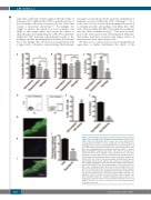
ABC
DEF
G
H
I
J
Figure 4. The meninges harbor quiescent and chemoresistant leukemia cells in vivo. (A-C) Mice were transplanted with NALM-6 leukemia cells (2x10⁶ cells; n=5 per group). After 4 weeks, mice were injected intra- venously with Click-iT EdU cell cycle reagent prior to harvesting tissues (meninges, bone marrow, and peripheral blood) and assessing for cell cycle (A-B) and the proliferation marker Ki-67 (C) by flow cytometry. (D-F) Mice were transplanted with membrane dye-labeled (DiD) or control, unlabeled NALM-6 leukemia cells (2x10⁶ cells) and then euthanized 4 weeks later (n=5). Ki-67 negative, quiescent (dye-positive) acute lym- phoblastic leukemia (ALL) cells were identified and quantified (D-F) in the meninges by flow cytometry. Representative flow cytometry plots are shown in (D). (G-I) Confocal microscopy images showing total (G; green), dye-retaining, quiescent (H; purple), and overlay (I) of leukemia cells detected in the meninges of mice 4 weeks after transplantation with dye- labeled (DiR) NALM-6 cells also expressing green fluorescent protein. (J) Mice were transplanted with dye-labeled human NALM-6 ALL cells (2x10⁶; n=5 per group). After 3 weeks, the dye-positive and dye-negative ALL cells were measured and quantified in the meninges and bone mar- row by flow cytometry before and after treatment with cytarabine 50 mg/kg for 3 days. A relative increase in the dye-positive leukemia cells after cytarabine treatment is consistent with dye-positive leukemia cells being chemoresistant relative to the dye-negative leukemia cells. For all graphs, *P<0.05, **P<0.01, ***P<0.001, ****P<0.0001 by analysis of variance or a t-test.
2136
haematologica | 2020; 105(8)
