
Page 158 - 2020_08-Haematologica-web
P. 158
L.M. Jonart et al.
weight (increasing its likelihood of CNS penetration), tol- erability in mice, lack of prior testing in leukemia, and likely multifactorial mechanism of action.37,38 Me6TREN significantly disrupted the adhesion of both NALM-6 and Jurkat leukemia cells to primary meningeal cells in a dose- dependent fashion (Figure 6A and Online Supplementary Figure S6B, C). This effect of Me6TREN on adhesion was not due to toxicity to leukemia or meningeal cells (Online Supplementary Figure S6D). We then quantified non-adher- ent leukemia cells in the cerebrospinal fluid of xenotrans- planted mice after treatment with either Me6TREN or phosphate-buffered saline control in order to measure dis-
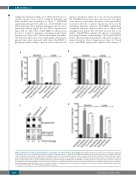
A
ruption of leukemia adhesion in vivo. As shown in Figure 6B, Me6TREN-treated mice showed a modest, but signif- icant, increase in leukemia cells in the cerebrospinal fluid, consistent with the co-culture experiment. Moreover, by disrupting leukemia adhesion, Me6TREN significantly attenuated leukemia chemoresistance in co-culture with meningeal cells (Figure 6C). We next assessed the in vivo ability of Me6TREN to enhance the efficacy of cytarabine in treating leukemia in the meninges. We tested NALM-6, Jurkat, and primary B-ALL leukemia cells with dosing reg- imens shown in Online Supplementary Figure S7. In all cases, Me6TREN significantly enhanced the efficacy of
C
D
Figure 2. Meningeal cells tilt the apoptotic balance of leukemia cells toward survival. (A, B) NALM-6 and Jurkat leukemia cells cultured in suspension or adherent to meningeal cells were treated with cytarabine 500 nM for 48 h and caspase-7 activity (A) and TMRE staining (B) assessed by flow cytometry. (C) NALM-6 leukemia cells were grown in suspension or adherent to meningeal cells for 48 h. Acute lymphocytic leukemia cells were then isolated and lysed. Protein lysate was used to probe a Human Apoptosis Antibody Array (Abcam). Representative portions of the membrane are shown. Relative protein expression was calculated after normal- ization using the IgG positive control. (D) BH3 profiling was performed using the BIM peptide on NALM-6 and Jurkat leukemia cells grown in suspension or adherent to meningeal cells for 24 h (NALM-6) or 48 h (Jurkat). Cytochrome c retention was measured by flow cytometry. Alamethicin is a peptide antibiotic that permeabilizes the mitochondrial membrane and serves as a positive control. For all graphs, data are the mean ± standard error of mean from three independent experiments. **P<0.01, ***P<0.001, ****P<0.0001 by analysis of variance.
B
2134
haematologica | 2020; 105(8)
